

 Blog
BlogCold Case - Part 1: The Cold Case Files
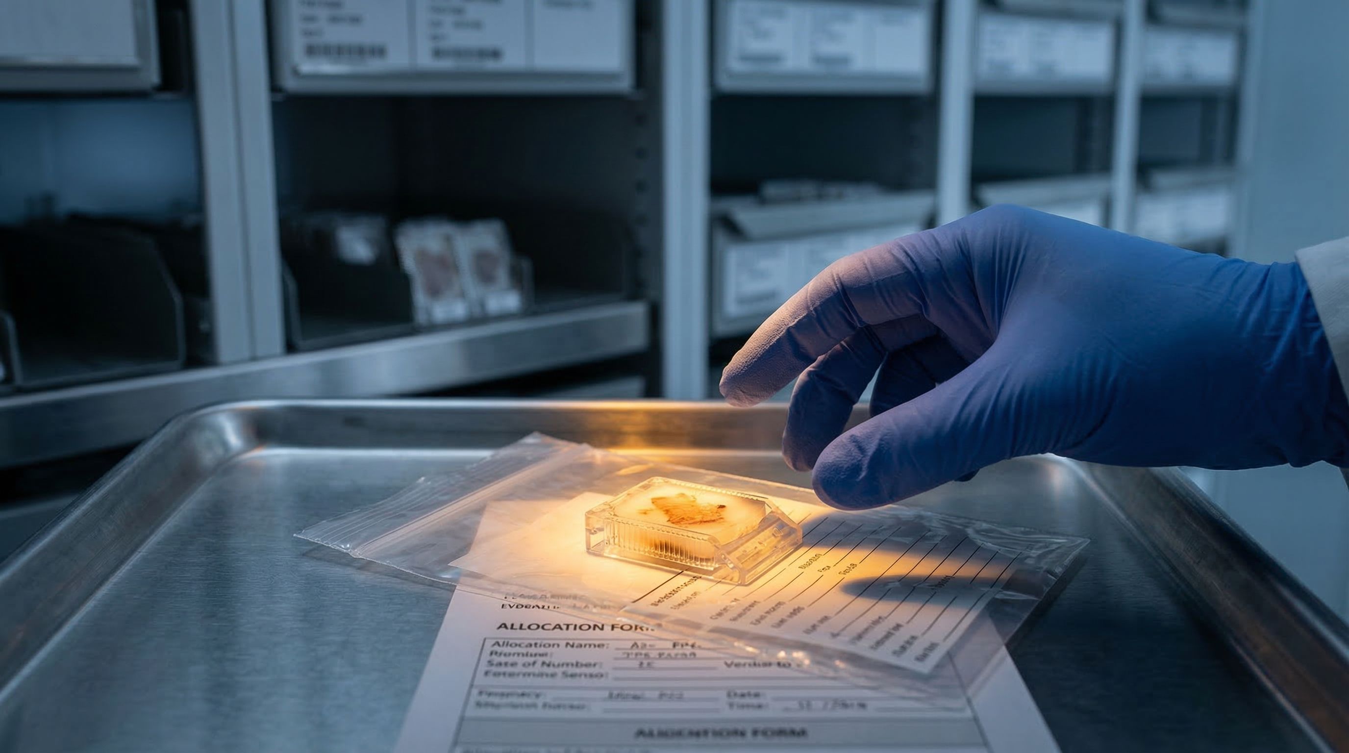
Millions of FFPE brain tissue blocks sit in biobanks worldwide, holding decades of evidence about Alzheimer's, Parkinson's, and neurodegenerative diseases. What if we could reopen these cases?
Blogs, publications, field guides, ebooks, application notes, and protocols for advanced cell analysis and automated tissue dissociation.
BlogMillions of FFPE brain tissue blocks sit in biobanks worldwide, holding decades of evidence about Alzheimer's, Parkinson's, and neurodegenerative diseases. What if we could reopen these cases?
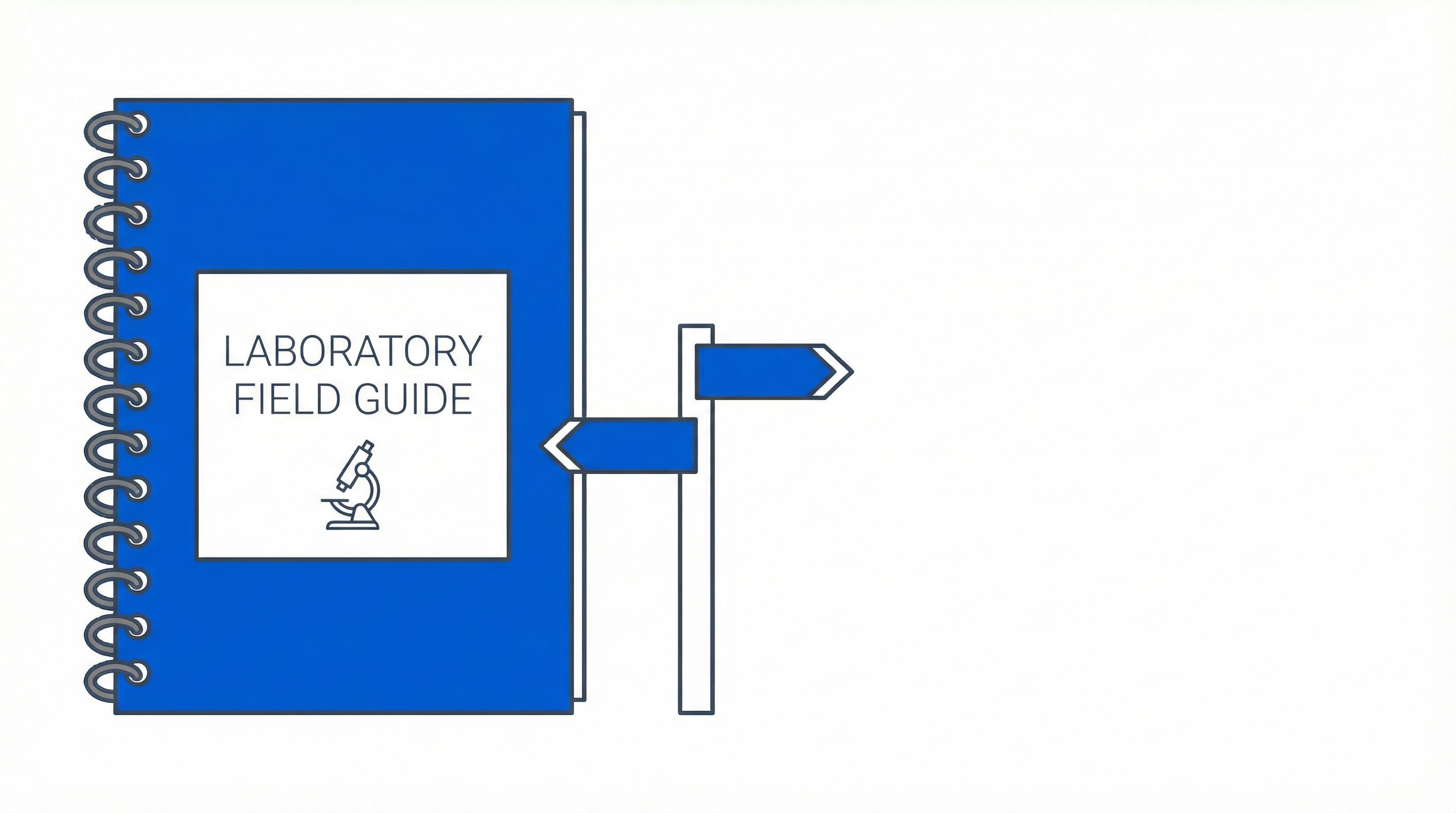 Field Guide
Practical strategies for maximizing nuclei recovery from limited FFPE tissue on the Singulator 200+, covering block quality assessment, sectioning waste reduction, the pilot curl approach for irreplaceable specimens, handling needle biopsies and crumbly blocks, and preserving nuclei yield post-processing.
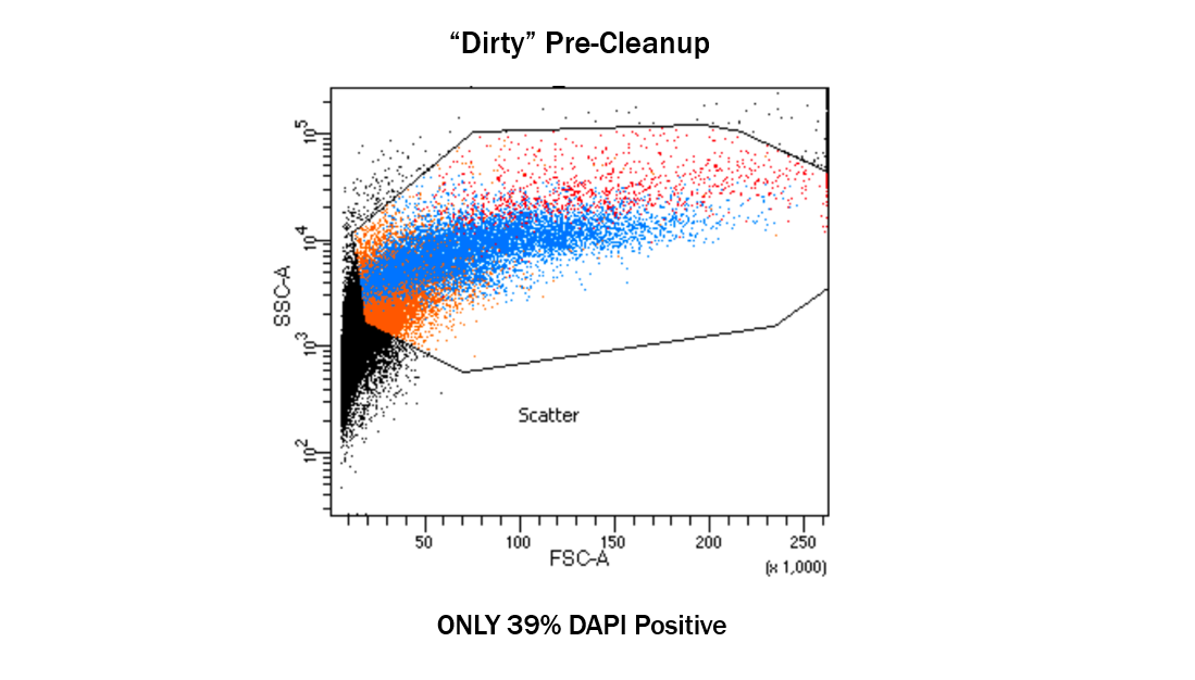
Field GuideQuality assessment of nuclei isolated from FFPE tissue using the Singulator 200+, covering yield measurement, DAPI staining for morphology, DV200 RNA quality metrics, erythrocyte contamination assessment, and structured go/no-go decision frameworks before downstream sequencing.
Field GuideEvery cell preparation contains some level of debris—fragments from lysed cells, extracellular matrix remnants, aggregates, and other particulate matter—that quietly skews counts and downstream results.
Field GuideEvery hour spent optimizing viability dye concentrations is an hour not spent on your actual experiments. It's optimized for use - that's the important thing. You don't have to optimize it as a customer. Pre-optimized viability reagents eliminate the titration experiments, the incubation testing, the cell-type-specific protocol development. Why spend time optimizing when validated performance is available from the first use?
Field GuideMost of the issue with viability reagents is that most people don't even know they exist. If you're running viability assays on Moxi V or Moxi GO II with generic dyes you optimized yourself, there's a better option you may not have heard about: pre-optimized, ready-to-use viability reagents designed specifically for your instrument. Now you know.
Field GuideSingle-cell genomics platforms capture transcripts from individual cells to reveal heterogeneity that bulk methods miss. That resolution depends on clean single-cell suspensions, where ambient RNA from lysed cells can contaminate every droplet.
Field GuideConsider a simple 1:100 dilution from stock. Errors can enter at multiple points—stock measurement, diluent measurement, mixing adequacy, transfer losses—and each step compounds into measurable variability.
Field GuideMost researchers assume that adding viability stains improves the accuracy of their cell counts. After all, you're adding more information—shouldn't that make the measurement better? The reality is more complicated.
Field GuideSearching for viability protocols, adapting literature methods, trial-and-error until something works - this is time you don't need to spend. The user manual is designed to be super easy. Concentration-based instructions tell you exactly what to do: put X amount of viability reagent and put X amount of sample, incubate and go. No protocol hunting required.
Field GuideA protocol can achieve high cell yield while producing low cell viability. Aggressive dissociation that frees many cells may simultaneously damage or kill them—a blindspot that viability assessment exposes.
Field GuideThat's why people - including me - just buy premixed gel loading dye and don't make my own from powder like my PI wanted me to. The same logic applies to viability reagents. Buy the damn gels rather than making them - consistency and data. When convenience and consistency matter more than tradition, pre-made beats DIY.
Field GuideEvery laboratory has protocols for cell culture, staining, and instrument operation. But ask about sample quality standards—specifically, debris thresholds—and you'll often find a blind spot where a QC checkpoint should be.
Field GuideUsing an aperture much larger than necessary reduces sizing resolution by creating smaller signal differences between cell sizes. Cell populations that should be distinguishable appear merged when the aperture is too large for the cells being measured. Target 15-40% of aperture diameter for optimal resolution. If you're counting lymphocytes on M+ cassettes because it works, you're sacrificing the sizing resolution that S+ cassettes would provide.
Field GuideWhen you make your own viability reagents, every batch is different. When you buy pre-optimized reagents with QC'd lot consistency, every lot performs the same. Long-term experiments need long-term consistency - and that consistency comes from manufacturing quality control, not from hoping your technique stays identical over months of work.
Field GuideThe 15 μm boundary provides clear selection criterion: cells under 15 micrometers use S+ cassettes, cells over 15 micrometers use M+ cassettes. This boundary isn't arbitrary - it's where each aperture size achieves the optimal 15-40% cell-to-aperture ratio for signal quality and sizing resolution. Know your cell size, follow the boundary, and cassette selection becomes automatic.
Field GuideEvery AI-based image counter was trained on a specific dataset, learning to recognize "cell" and "not cell" from images someone curated. When your sample doesn't match that training set, physics-based counting tells a more reliable story.
Field GuideThe Precision Cell Systems Singulator provides automated, standardized tissue dissociation. But automation doesn't mean zero debris or guaranteed sample quality—integrating a Moxi QC checkpoint is what makes the workflow dependable.
Field GuideSingle-cell genomics workflows have a critical decision point: do you load this sample onto an expensive chip, or does it need more cleanup first? A preloading QC checkpoint answers that with confidence.
Field GuideTIL counting and immune cell killing assays are immediate applications for dual-cassette workflows. Cancer cells are large, T cells are small - no single cassette optimizes both. Run S+ for accurate T cell counts, M+ for accurate tumor/target counts. The extra run takes minutes but delivers publication-quality E:T ratios and killing percentages.
Field GuideNew lab members need to generate valid data quickly. Teaching protocol optimization takes weeks. Teaching protocol execution takes minutes. The user manual is designed to be super easy - put X amount of viability reagent, put X amount of sample, incubate and go. When reagents are pre-optimized, training focuses on execution, not development.
Field GuideSmall cells measured through oversized apertures generate weak electrical signals that fall below detection thresholds or get confused with debris. If you're counting lymphocytes, PBMCs, Jurkat cells, or any suspension lines under 15 micrometers with the wrong cassette, you're likely undercounting. Switch to S+ cassettes where the smaller aperture ensures your small cells generate strong, detectable signals clearly distinguishable from noise.
Field GuideLarge cells approaching the aperture diameter create artificially high signals and risk clogging the sensing orifice. Clogging interrupts runs, wastes samples, and requires cassette replacement mid-experiment. For adherent cell lines like CHO, HEK293, and HeLa, and for primary tissue cells over 15 micrometers, M+ cassettes provide the larger aperture necessary to prevent physical blockage.
Field GuideWhen your sample contains both small and large cells, no single cassette optimizes measurement for both populations. The solution: run the same sample twice - once with S+ to get accurate small cell counts, once with M+ to get accurate large cell counts. This dual-cassette workflow delivers accurate data for both populations rather than compromised data for everyone.
Field GuideCoincidence - multiple cells in the aperture simultaneously - causes two cells to be counted as one, corrupting both count and size data. Optimal aperture utilization means targeting 15-40% of aperture diameter so cells generate strong signals while avoiding coincidence artifacts. Match your cassette to your cell size, stay within concentration guidelines, and coincidence becomes a non-issue.
Field GuideBrain FFPE tissue creates unique nuclei isolation challenges. Myelin debris, lipid contamination, and fragile neuronal nuclei require controlled automated processing to preserve cell-type diversity for single-nucleus sequencing.
Field GuideProcess brain tumor FFPE from surgical resections on the Singulator 200+. Preserve cancer cells and immune populations for snRNA-seq and spatial analysis.
 Ebook
EbookHow the Singulator 200+ preserves fragile neuronal nuclei from irreplaceable postmortem brain tissue. Automated FFPE processing for Alzheimer's, brain tumors, and brain atlases.
Field GuideExtract nuclei from FFPE brain tissue for Alzheimer's, Parkinson's, and Lewy body research. Longitudinal cohorts, cell-type preservation, and disease staging on the Singulator 200+.
Field GuidePair spatial transcriptomics with snRNA-seq from the same FFPE brain block. Block allocation, platform selection, and nuclei quality for multi-omic brain studies.
Field GuideGet high-quality nuclei from limited postmortem brain FFPE sections. Process NIH biobank allocations, hospital archives, and surgical specimens on the Singulator 200+.
Field GuidePractical guide to postmortem brain FFPE challenges: necropsy timing, fixation variability, biobank sourcing, myelin debris, and how the Singulator 200+ standardizes nuclei extraction.
Field GuideStandardize brain FFPE nuclei extraction across consortium sites with the Singulator 200+. Eliminate operator variability, prevent batch effects, and scale for atlas projects.
 Webinar
Ronan Chaugnet from Memorial Sloan Kettering shares several years of head-to-head benchmarking data comparing nuclei extraction platforms for FFPE tissue. The Singulator 200+ and Miltenyi GentleMACS both outperformed manual approaches across glioblastoma, liver, lung, pancreas, and tumor samples, with the Singulator 200+ standing out for its fully automated, enzyme-free workflow. Ronan also walks through three methods his lab has built on top of automated FFPE extraction: 10X Genomics FLEX for high-quality gene expression, PERFF-Seq for sorting rare cell populations by RNA markers, and GIFT-Seq for detecting 600+ mutations at single-cell resolution from archival tissue.
 Protocol
This protocol describes how to isolate, count, and prepare single nuclei from FFPE (formalin-fixed, paraffin-embedded) tissues for single-nuclei sequencing assays using the Singulator 200+ platform. Deparaffinization and rehydration of the tissue using solvent and various concentrations of ethanol are conducted on the Singulator 200+ platform along with automated nuclei isolation.
ProtocolNuclei counting protocol for Moxi V
ProtocolViability staining protocol for Moxi V
ProtocolValidation check protocol for Moxi V
ProtocolWBC Counting Protocol for Moxi Z
ProtocolThis protocol describes an approach to achieve rapid, highly-accurate nuclei / nucleated-cell counts by first lysing the cells to isolate the nuclei, labeling the nuclei with PCS Viability Reagent, and counting using the Moxi GO II system
ProtocolSystem Check Bead Protocol for Moxi Z
ProtocolThis protocol describes how to isolate, count, and prepare single nuclei from FFPE (formalin-fixed,paraffin-embedded) tissues for single-nuclei sequencing assays using the Singulator Platform.
ProtocolThis protocol describes how to stain with both propidium iodide and acridine orange. It also explains how to use compensation settings on the Moxi GO II
ProtocolThis protocol outlines a standardized dual-staining workflow for the Moxi GO II to simultaneously quantify apoptosis and viability using FITC-Annexin V and concentrated PCS Viability Reagent.
ProtocolThis protocol outlines a dual-staining procedure using Calcein-AM and Propidium Iodide on the Moxi GO II to simultaneously assess cellular membrane integrity and metabolic vitality for a comprehensive evaluation of cell health.
ProtocolThis protocol details the standardized preparation, blocking, and immunolabeling of single-cell suspensions using either direct or secondary antibody staining workflows optimized for high-resolution population analysis on the Moxi GO II flow cytometer.
ProtocolThis protocol outlines the process for isolating, cleaning, counting, and preparing single cells fromfresh intestine tissue for single-cell sequencing assays listed below. Some
ProtocolThis protocol outlines the process for isolating, cleaning, counting, and preparing single cells from fresh tissue for the single-cell sequencing assays listed below
ProtocolThis protocol outlines the steps for isolating, counting, and preparing single cells from FFPE (formalin-fixed,paraffin-embedded) or paraformaldehyde (PFA) fixed whole tissues for single-cell sequencing assays using theSingulator™ Platform. The process includes automated deparaffinization, rehydration, and dissociation.
ProtocolThis protocol describes how to isolate, clean, count, and prepare single nuclei from fresh frozenmouse brain tissue for single-nuclei sequencing applications. Some optimizations of theSingulator™ protocol parameters may be needed based on storage time and tissue condition.
ProtocolThis protocol describes how to isolate, clean, count, and preparesingle nuclei from frozen tissue for single-nuclei sequencing assays.
ProtocolThis protocol provides step-by-step instructions for preparing, running, and gating validation check beads to verify the sizing, concentration, and fluorescence accuracy of the Moxi GO II instrument
ProtocolThis document provides a detailed protocol for isolating, stimulating, fixing, permeabilizing, and intracellularly immunolabeling white blood cells from whole blood samples for analysis on the Moxi GO II.
ProtocolThis document provides a step-by-step protocol for measuring oxidative stress in living cells by detecting reactive oxygen species (ROS) using the CellROX Green assay on the Moxi GO II.
ProtocolThis document provides a step-by-step protocol for immunolabeling the surface of white blood cells while effectively eliminating red blood cell interference through lysis and optional buffy coat isolation for analysis on the Moxi GO II.
 BlogPart 5
Brain atlases are being built. Archival tissue is finally talking. How standardized nuclei extraction and platform-agnostic analysis are solving neuroscience's oldest cold cases.
 BlogPart 2
Manual extraction of nuclei from FFPE brain tissue destroys 50 to 60 percent of starting material. Fragile neurons die first, leaving biased results. Here is what goes wrong.
BlogPart 1Millions of FFPE brain tissue blocks sit in biobanks worldwide, holding decades of evidence about Alzheimer's, Parkinson's, and neurodegenerative diseases. What if we could reopen these cases?
 BlogPart 4
Manual FFPE processing destroys fragile neuronal nuclei and produces variable results. The Singulator 200+ automates the workflow with a two-cartridge system that delivers consistent, operator-independent results from irreplaceable brain tissue.
 BlogPart 3
NIH biobanks give you one allocation of irreplaceable brain tissue. Manual processing destroys 50-60% before analysis begins. The extraction method is the real variable.
Field GuideEnd-to-end protocol walkthrough from FFPE block selection through deparaffinization, nuclei isolation, quality control, library preparation, sequencing, and data analysis using the Singulator 200+.
Field GuideHow the Singulator 200+ GREEN cartridge automates FFPE deparaffinization and rehydration, eliminating toxic solvents, fume hoods, and manual ethanol series from nuclei extraction workflows.
Field GuidePractical guide to connecting Singulator 200+ FFPE nuclei with downstream analysis platforms including 10x Chromium Flex, Xenium, ATAC-seq, Visium, and MERFISH, covering quality requirements, expected yields, and multi-platform study design.
Field GuidePractical guide to preparing FFPE tissue inputs for the Singulator 200+ automated nuclei extraction platform, covering curl thickness selection, tissue mass requirements, block age effects, quality assessment, and handling difficult or precious specimens.
Field GuidePractical troubleshooting guide for the Singulator 200+ FFPE nuclei extraction workflow, covering the five most common problem categories: low yield, excessive debris, poor RNA quality, batch-to-batch variability, and cartridge/instrument errors.
Field GuidePractical strategies for maximizing nuclei recovery from limited FFPE tissue on the Singulator 200+, covering block quality assessment, sectioning waste reduction, the pilot curl approach for irreplaceable specimens, handling needle biopsies and crumbly blocks, and preserving nuclei yield post-processing.
Field GuideQuality assessment of nuclei isolated from FFPE tissue using the Singulator 200+, covering yield measurement, DAPI staining for morphology, DV200 RNA quality metrics, erythrocyte contamination assessment, and structured go/no-go decision frameworks before downstream sequencing.
 Ebook
EbookThe most comprehensive tissue dissociation reference available. Interactive protocols for single-cell isolation and nuclei extraction across 57+ tissue types with community and Singulator-optimized methods.
EbookHow formalin-fixed paraffin-embedded tissue archives hold enormous potential for single-cell genomics, and how the Singulator 200+ automates the nuclei extraction workflow to unlock that potential
 Ebook
EbookThe debris problem: Membrane fragments, aggregates, media residue, and lysed cell debris are present in virtually every biological preparation. How does your counting method distinguish a cell from a piece of debris?
 Publication
In this study, the authors used a Moxi Z Cell Counter to count trypsinized, treated PANC-1 cells so they could re-plate defined cell numbers for colony formation assays and accurately calculate plating efficiency and survival fraction after PAA-TiOx nanoparticle and X-ray treatments under normoxic and hypoxic conditions.
PublicationThe authors used a Moxi Z to count cells as part of preparing mammalian cell cultures for in vitro nanovanilloid testing (viability/oxidative-stress assays and TRPV1 calcium-influx imaging).
PublicationIn this study, the authors used the Moxi GO II to quantify endothelial apoptosis after Hif2α siRNA knockdown and hypoxic exposure, using Annexin V–FITC and PI with the instrument’s apoptosis app to measure cell-death outcomes under their ischemia-relevant conditions.
PublicationIn your staged partial-dissociation retinal organoid workflow, you used the Moxi Go II to count the filtered dissociation-fraction cell suspensions immediately before 10x Chromium X scRNA-seq loading (targeting ~8,000 cells per sample), enabling consistent single-cell capture across fractions and lines.
PublicationThe authors used a Moxi V to measure Aspergillus niger spore concentration, enabling per-10^6-spore normalization of downstream plate-reader GFP fluorescence when comparing transformants across binary vector ORI variants.
PublicationThe authors used the MOXI Z Mini to count and standardize 50,000 NIH3T3 cells per condition immediately before ATAC-seq library preparation, supporting their chromatin-accessibility comparisons at the Actg2 locus across RNA-destabilizing perturbations.
PublicationThe authors used a Moxi Z to count isolated bovine PBMCs after density separation and washes, then normalized each sample to ~2×10^7 cells per tube prior to RNAlater stabilization and downstream bulk RNA-Seq.
PublicationThe author used a platelet–fibroblast co-culture model, and a Moxi-Z Coulter-based cell counter to quantify RPC-C2A fibroblast cell numbers after UVC injury and platelet/lysate treatment and to normalize fibroblast-associated ATP measurements to cell count, supporting your survival and bioenergetic conclusions.
PublicationThe authors used a Moxi GO II to perform fluorescence-based flow cytometric phenotyping of BM-derived cultures (CD45 and intracellular RUNX2/BAP) to verify the osteoblastic differentiation context for interpreting downstream α-radiation–responsive omics measurements.
PublicationIn this study, the authors used the Moxi GO II to run a FITC-based apoptosis assay on treated prostate cancer cells (±5-ALA, ±irradiation) and quantify early apoptotic fractions as a mechanistic readout of radiosensitization.
PublicationThe authors used a Moxi cell counter (Orflo, S cassettes) to quantify the live, post-FACS-sorted MOE single-cell suspension and dilute it to ~1000 cells/µL for 10x Genomics single-cell RNA-seq loading.
PublicationIn this study, the authors used a Moxi Z to quantify H. akashiwo cell-density changes in phosphate-depleted co-cultures and a Moxi Go II Mini to measure CellTracker™ Green–labeled bacterial fluorescence associated with algal-sized, red-autofluorescent particles as evidence of bacterivory linked to growth rescue under phosphate limitation.
PublicationIn this study of Naegleria’s mitotic vs differentiation microtubule programs, the authors used a Moxi Z impedance-based counter to quantify amoeba concentration so they could inoculate and grow cultures at defined densities before initiating the mitotic synchronization workflow.
PublicationIn this study, the authors used a Moxi GO II (561 nm LP filter) to quantify TMRM fluorescence in PBMCs and calculate inhibitor-dependent shifts in the fraction of TMRM-positive cells as a small-sample, intact-cell readout of mitochondrial membrane potential (∆Ѱm) and Complex V operating mode.
PublicationThe authors used an ORFLO Moxi Mini to count viable cells after irradiated MRC5-hTERT and HCT116 pellets were re-plated and expanded for 5–6 days, using the averaged viable-cell counts (normalized to same-day non-irradiated controls) as the viability endpoint to compare inter-pulse timing conditions.
PublicationThe authors used a Moxi Z to count trypsinized vascular smooth muscle cells after a 3-day antiviral treatment, providing the primary quantitative endpoint for proliferation in their remdesivir vs. molnupiravir/nirmatrelvir comparisons.
PublicationThe authors used a Moxi Z (Type-M cassettes) to collect 24-hour interval EO771 cell counts and build proliferation curves to test whether BMP9 treatment or BMPR2-silenced endothelial conditioned media/ECM altered EO771 growth.
PublicationIn this study, the authors used a Moxi GO II with propidium iodide to measure cell count and viability of αTC cells after Klf4 siRNA transfection, supporting the subsequent qPCR-based knockdown validation and selection of knockdown samples for downstream mechanistic assays.
PublicationIn this TNBC cisplatin in vitro study, the authors used the Moxi GO II to run an Annexin V (FITC) / PI apoptosis flow-cytometry assay (and to capture 24-hour flow-based cell-count outcomes) to quantitatively compare treatment-induced killing in TNBC versus epithelial cells across mono-culture and co-culture conditions.
PublicationThe authors used a Moxi GO II to count and monitor CAR T cells (including cell size/growth kinetics) throughout expansion, advancing the products into in vitro cytotoxicity assays and in vivo PTCL PDX studies only once the cells’ kinetics/size indicated they had “rested” from stimulation.
PublicationThe authors used a Moxi cell counter to accurately seed 10,000 iPSCs per well when generating human cardiac organoids, ensuring consistent starting organoid inputs before assessing fluorescent nanoparticle biodistribution and cell-type–specific uptake by downstream fluorescence microscopy.
PublicationThe authors used a Moxi Z to count primary pulmonary artery endothelial cells after trypsinization—both to seed 6000 cells/well and to perform serial 24-hour cell counts through 120 hours—to quantify and compare PAEC proliferation between control and Glenn lambs.
PublicationThe authors detached IMR-90 fibroblasts with trypsin and used a Moxi V cell analyzer to count the cells, with no further stated use of the Moxi readout for secretome normalization in the proteomics workflow.
PublicationThe authors used the Moxi GO II to measure cell concentration and ensure optimal loading density when preparing neonatal umbilical cord blood single-cell suspensions for 10× Genomics Chromium scRNA-seq.cytometer (Orflo Technologies) to ensure optimal loading density.
PublicationThe authors used a Moxi GO II to count engineered T-cell cultures every other day and monitor cell size/growth kinetics as a practical criterion for when the cells had rested from stimulation and were ready to proceed into downstream adoptive T-cell experiments.
PublicationThe authors used a Moxi Go II to verify post-thaw PBMC viability and accurately count cells so they could plate a consistent 1×10⁶ cells per condition for HIV Gag/Env peptide stimulation prior to intracellular cytokine staining and polyfunctionality analysis by flow cytometry.
PublicationThe authors used the Moxi GO II to measure PE-labeled cell-surface Nectin-4 expression by flow cytometry in a rectal cancer cell line, providing protein-level confirmation of Nectin-4 knockdown used to interpret downstream proliferation/5-FU response assays.
PublicationIn this study, the authors used a Moxi Z to count human airway smooth muscle cells 72 hours after miRNA mimic transfection, enabling them to quantify relative proliferation vs scramble control as functional validation for asthma- and rhinitis-associated cord-blood miRNA signatures.
PublicationThe authors used a Moxi Z to count CD14+ bovine monocytes after magnetic enrichment and standardize seeding concentration prior to ex vivo differentiation and downstream antigen-stimulation readouts (MAPK ELISA and RNA-seq).
PublicationIn this scoliosis wound-healing study, the authors used a Moxi Z to count cells recovered from POD1 surgical wound drainage after RBC lysis, then used that counted cell suspension as the input for multicolor flow-cytometry immunophenotyping to compare wound-associated leukocyte populations between idiopathic and neuromuscular patient groups.
PublicationIn this study, the authors used the Moxi Go II to quantify lysosomal activation in Lysosomal-METRIQ reporter dermal fibroblasts by measuring GFP and RFP fluorescence and computing a GREEN/RED ratio after resveratrol (and control drug) treatments, providing functional evidence that resveratrol enhances lysosome-dependent degradation pathways.
PublicationThe authors used a Moxi cell counter to quantify neutrophil chemotaxis by counting the number of cells that migrated through a 3-µm Boyden-chamber membrane into the lower wells after 90 minutes of fMLF-driven migration (with or without BLI pretreatment).
PublicationThe authors used a Moxi GO II to measure viability and concentration of dissociated monarch brain single-cell suspensions immediately before 10X Genomics single-cell capture for scRNA-seq.
 App Note
Much of the hassle of analyzing PBMC samples comes from the inadequate counting of the cells of interest, usually because of debris or lack of robust counting. By circumventing imaging and going back to gold-standard physics-based principles, every Moxi instrument gives unparalleled abilities in getting the best cell count. With the added opportunities for fluorescence-based detection with a Moxi V and Moxi GO II, PBMC viability checks have never been more accurate. Ensure your precious samples aren’t wasted by making sure you get the quality checks right the first time by relying on a Moxi instrument.
App NoteScientists are concerned with speed, accuracy, and convenience, and those running the lab and industries are concerned with the cost typically associated with high-performing instruments. Our proprietary Coulter Principle-based system delivers on all three accounts, and is therefore a perfect fit for any cell biology benchtop.
App NoteThere are a variety of ways to monitor this process in cell culture, the two most prominent being cell counting via imaging-based and coulter-based cell counting methods and higher-level cellular analysis with fluorophore-tagged antibodies and fluorescent reporters via flow cytometry. While each instrument used to achieve these two things has its advantages and disadvantages, few and far between can do both at once. Out of all your options, no other instrument has the power to do both with the speed, precision, and ease that Precision Cell Systems' Moxi Go II can. In this document, we will take you through how you can unlock the next stage in your cellular analysis process for CAR-T with our instruments.
App NoteProtocols for single-cell sequencing library preparation present several challenges along the way that can impact the accuracy and quality of downstream sequencing results. Making sure you overcome these is essential for not wasting valuable sample and money on failed sequencing runs. Precision Cell Systems’ Moxi line of Coulter Principle-based cell analyzers enable you to address all of these variables in a single instrument, making it the ideal go-to for all of your sequencing needs.
App NoteHere, we present the Precision Cell Systems Moxi GO II system as a simple, rapid, and effective flow cytometric approach to the study of apoptosis using Annexin V
App NoteThe triple-layered cell health assessment where the first layer of isolating cells from debris via event size, the second layer of PI for staining dead cells, and the third layer of AO for all cells or calcein for only live cells ensures that every event that is a real cell has been classified as alive or dead and none are left uncounted. The less layers that are used, the more unreliable the results. Multiple cell counters on the market are capable of doing this, but the accuracy and repeatability of data generated by a Moxi Go II makes it the clear winner.
App NoteThe Singulator platform paired with Parse Biosciences technology enables efficient single-cell isolation and sequencing from challenging tissue types, with high viability and robust cell yields suitable for downstream scRNA-seq analysis.
App NoteThese data demonstrate the ability of the Singulator Platform to prepare single-cell suspensions for both positive selection by a cell surface marker and single-cell RNA sequencing. These methods enable researchers to identify genes of interest for potential therapeutic targets for anti-tumor response
App NoteThe Singulator™ Platform provides a reproducible and precise method for isolating nuclei from complex tissues suitable for single-nuclei RNA sequencing. Our results demonstrate consistent nuclei yield and high-quality sequencing metrics across biological replicates, making it a reliable tool for neuroscientific research. The ability to produce high-quality nuclei with minimal variability enhances the accuracy of downstream snRNA analyses, facilitating deeper insights into normal biology and disease. Finally, the ability to identify rare cell types underscores the efficacy of the Singulator Platform in comprehensive cellular analysis, providing a detailed representation of the cellular landscape.
App NoteThe presence of intracellular and extracellular debris in single-nucleus RNA sequencing can present a significant challenge in obtaining reliable and accurate results. However, the use of the Precision Cell Systems Nuclei Debris Removal Reagent can effectively improve the quality of the samples. Our evaluation of the Nuclei Debris Removal Reagent shows successful removal of debris from traditionally high-debris samples of brain and liver nuclei while delivering high quality single nuclei suspensions. The resulting samples are able to be run through microfluidic single nuclei sequencing platforms without issues of clogging or reduced data quality
Field GuideAI segmentation algorithms fail when encountering debris, clusters, or samples they were not trained on—resulting in minimum 3-4% error per image even under.
 BlogPart 1
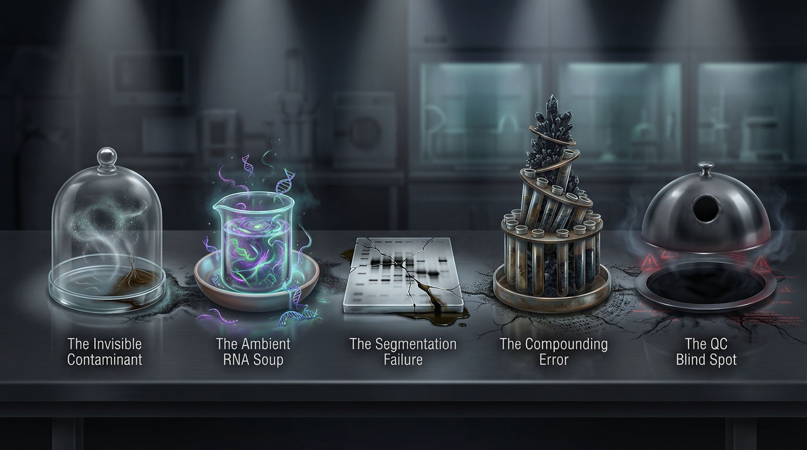
A cell count without debris quantification is like a menu without an ingredient list. Without knowing what invisible contaminants are present, every downstream decision becomes a gamble.
 BlogPart 2
Five contaminants corrupt every sample. Image counters exclude them from counts but never reveal their presence. Until you can quantify what's actually in your sample—not just how many cells—these villains control the menu.
 BlogPart 3
Better AI won't save your data. More training images won't expose the contaminants. Faster cameras won't quantify your debris. Physics will. The Coulter principle—the same physics that transformed clinical hematology—offers research laboratories what imaging never can: direct measurement of what's actually in your sample.
 BlogPart 4
The recipe for defeating the five villains isn't better algorithms or faster cameras. It's physics-based measurement that reveals what image counters hide: the complete composition of your sample. Direct size measurement. Complete population visualization. Standardized thresholds. Informed decisions. That's the recipe.
 BlogPart 5
Every laboratory has an invisible menu. Hidden ingredients contaminate samples. Villains corrupt data. Resources get wasted on samples that should have been cleaned up first. The question isn't whether these problems exist—they do, in every laboratory that relies on image-based counting alone. The question is whether you'll continue ordering blind, or finally demand to read the full ingredient list. Physics-based debris quantification isn't just an alternative to image counting. It's the missing QC checkpoint that transforms sample preparation from guesswork to measurement. From hope to confidence. From invisible menus to clean kitchens. The recipe is proven. The villains are defeated. The kitchen can be clean.
PublicationIn this study, the authors used Moxi to standardize plated cell numbers and per-reaction cell inputs for proliferation, neuronal differentiation phenotyping, and genome-wide CHD7 occupancy assays in iMOP progenitors and iMOP-derived neurons.
Field GuideEvery single-cell experiment represents a significant investment—consumables alone cost $500–1000+ per chip, plus downstream sequencing—so a confident go/no-go decision before chip commitment protects the run.
 BlogPart 1
Somewhere in your institution, there's a dusty shelf. Sitting on it: thousands of tissue blocks. Each one holds decades of clinical history. Patient outcomes. Treatment responses. Disease progression. Data that took years to collect. And can you blame researchers for walking past it?
 BlogPart 2
Here's a weird fact: the same chemical that saves your sample also traps everything useful inside it. Think about laminating a document. Great for protection. Terrible if you need to edit what's inside. That's formalin. And for decades, nobody could undo the damage.
 BlogPart 3
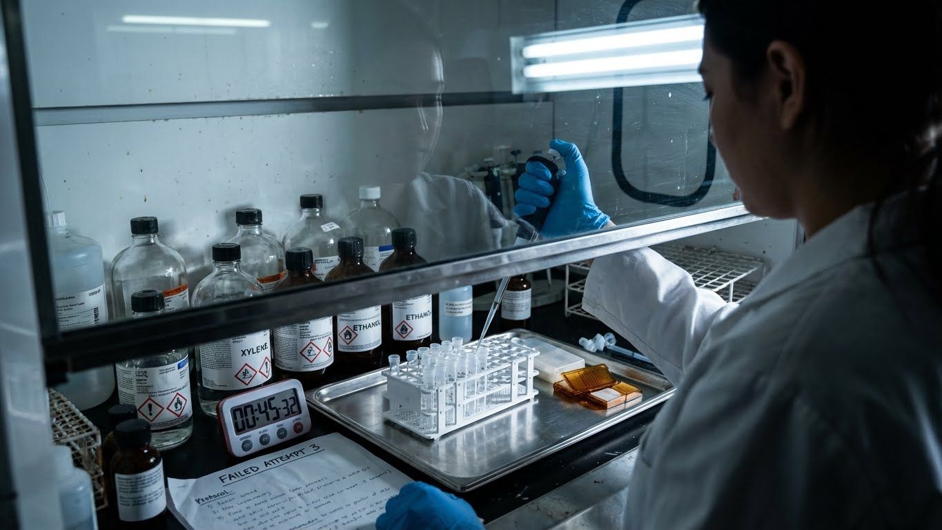
Third try. Third failure. The fume hood is running. The xylene smells terrible. And the protocol that worked last Tuesday is giving nothing but debris. Sound familiar? Here's the question nobody was asking: Is it your technique—or is it the physics?
 BlogPart 4
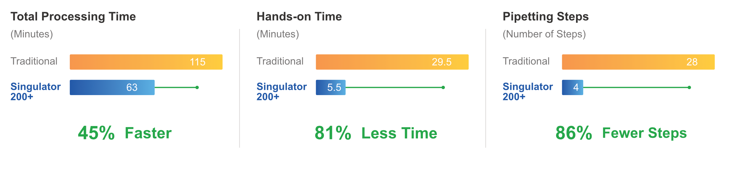
What if that two-hour protocol took sixty minutes? What if twelve pipetting steps dropped to four? What if the fume hood became optional? These aren't hypotheticals. This is what purpose-built automation looks like. And the difference isn't just time.
 BlogPart 5
Behind every tissue block is a patient who said yes. Yes to collection. Yes to research. Yes to the hope that their gift might help someone else. They trusted the science. The question now: are the protocols worthy of that trust?
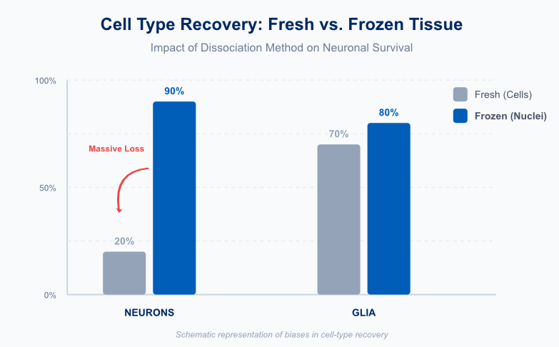 BlogPart 3
Fresh vs. Frozen: Which Side Are You On? Description: The debate between fresh tissue (whole cells) and frozen tissue (nuclei) divides labs. We explore why fresh dissociation often creates a "map of a disaster" through stress artifacts, and why frozen nuclei offer the unbiased, stable truth required for atlas-scale science.
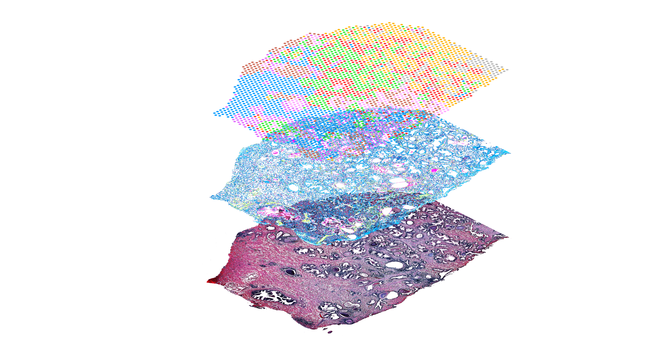 BlogPart 4
The List is Good, But Is the Map Better? Description: Single-nucleus sequencing gives you the "List" of cell types, but Spatial Transcriptomics gives you the "Map." Discover how combining these technologies creates a high-resolution "Precision Point," and how one automated platform can serve as the engine for both workflows.
 BlogPart 5
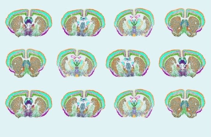
From the hidden world of glia to the final 3D atlas, the journey of modern neuroscience relies on one foundational step: sample preparation. We conclude our series by challenging researchers to prioritize clean, reproducible input to build the definitive map of the human brain.
 BlogPart 1
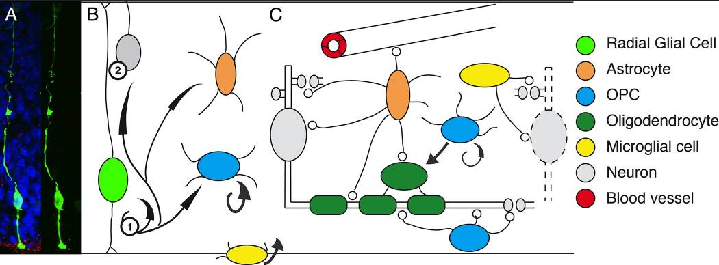
For a century, neuroscience focused exclusively on the neuron, dismissing glia as mere "packing peanuts." Discover how single-nucleus sequencing revealed the active, critical role of the brain's immune and support systems—and why this shift changes everything for Alzheimer's research.
 BlogPart 2
What is Ruining Your Frozen Experiments? Description: Every great story needs a villain. In frozen brain tissue processing, that villain is myelin debris. Learn how lipid contamination clogs microfluidics and ruins data, and see how the Singulator’s automated Protocol DP0006 neutralizes this threat to unlock biobank archives.
PublicationPublicationOur team is here to help you find the right resources for your research.
Contact Us