Cold Case - Part 1:
The Cold Case Files
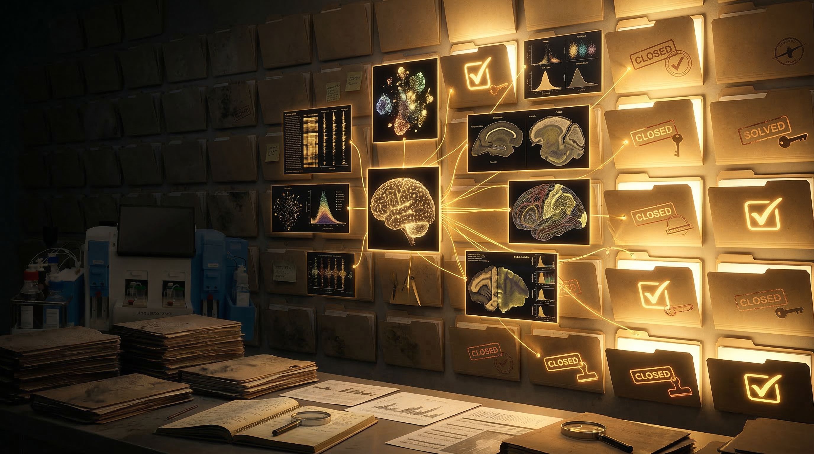
Cold Case: When Brain Tissue Holds the Evidence - Part 1 of 5
(Prefer to listen to the full story? Click play to hear Episode 1 of our accompanying audio version.)
The Precision Point - Cold Case - Episode 1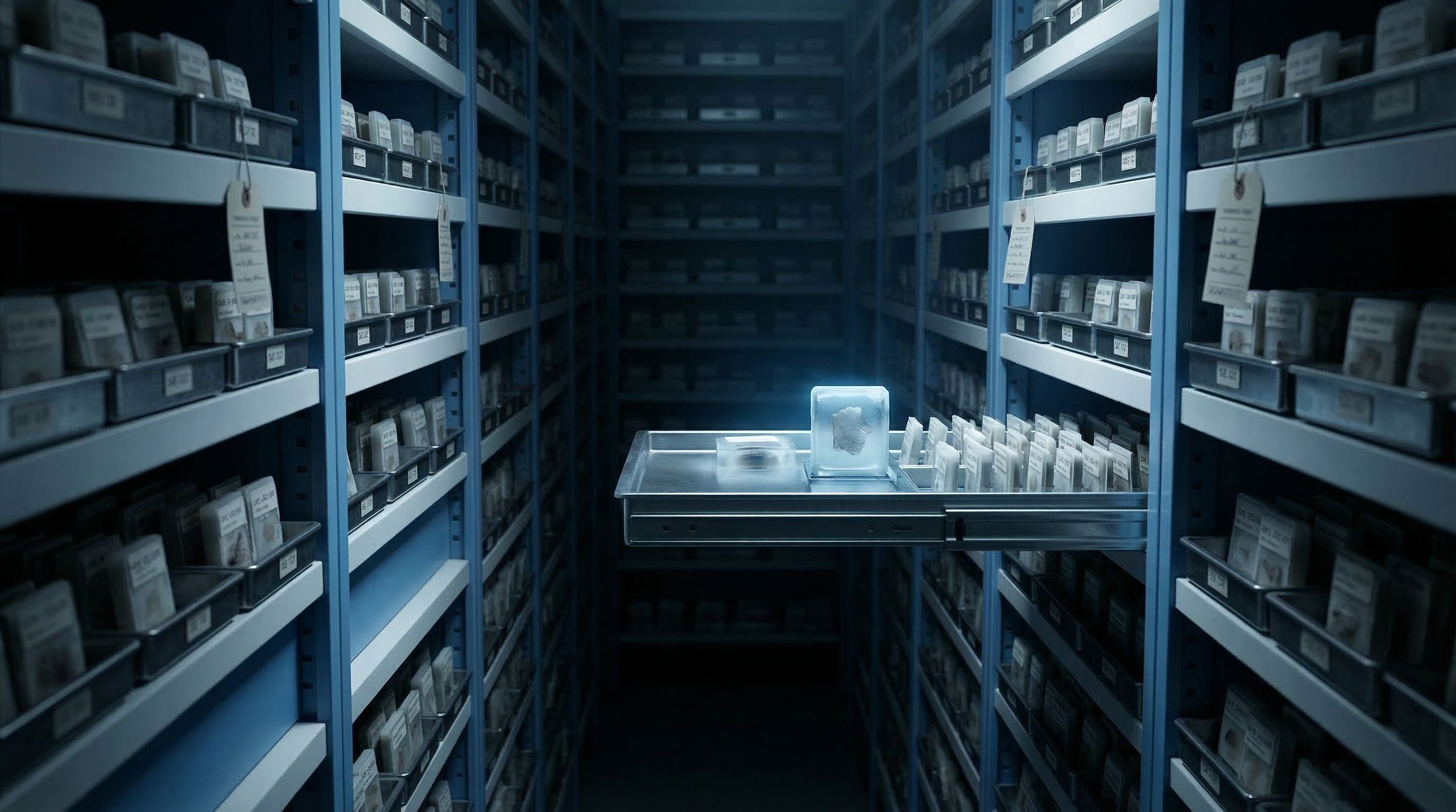
Somewhere in a hospital basement, behind a fireproof door and a keycard lock, rows of metal shelves hold more evidence about brain disease than any living researcher has ever examined.
The shelves are not marked with case numbers. They are labeled with patient identifiers, tissue codes, and dates that sometimes go back thirty years. What fills these shelves is not paper. It is paraffin, thousands of small white blocks the size of dice, each one holding a thin slice of someone's brain.
This is a biobank. And every block on these shelves is an unsolved case.
The evidence we already have
The numbers are staggering. Major brain banks around the world hold collections running into the hundreds of thousands of tissue blocks. The Harvard Brain Tissue Resource Center alone has collected over 10,000 brain donations. The UK Brain Bank Network holds samples from thousands more. Multiply that across medical centers, university pathology departments, and national tissue repositories, and the total runs into the millions.
Each block tells a story. An Alzheimer's patient followed for twenty years through cognitive testing, brain imaging, and clinical observation, whose brain was donated after death. A Parkinson's patient whose tremor started in her left hand at age 54 and progressed over a decade, with detailed neurological records at every stage. A rare Lewy body case where the family agreed to autopsy because the diagnosis was uncertain until the end.
The clinical histories are detailed. The tissue is preserved. The cases sit on shelves, waiting.
Why the cases went cold
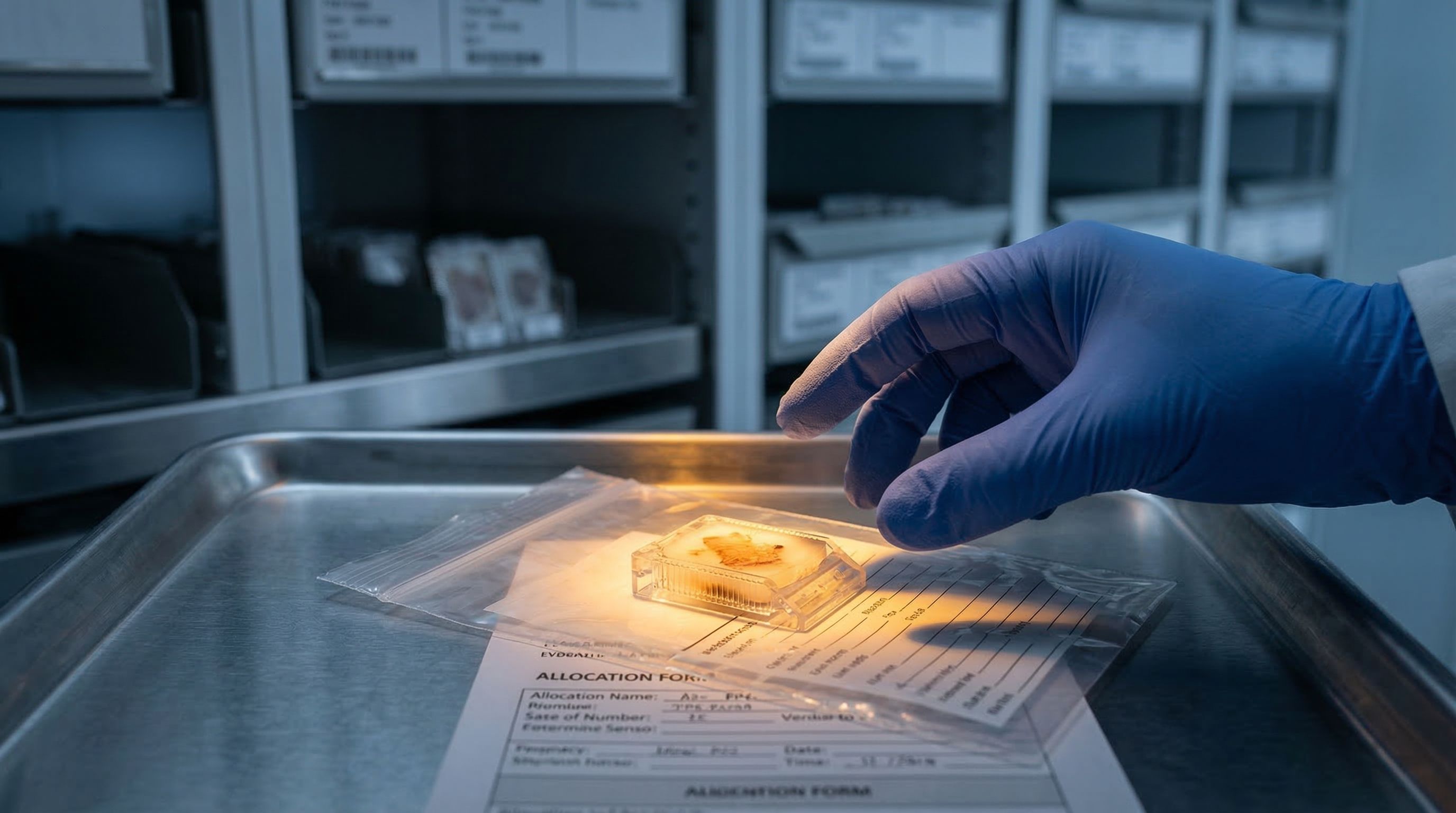
Brain tissue does not come easily to research. Every block in that biobank exists because someone died, and because that person or their family decided to donate. There is no going back for a second sample. What sits in paraffin is all there is.
For decades, pathologists could examine these samples under a microscope. They could stain for proteins and identify broad patterns of disease, the plaques of Alzheimer's, the Lewy bodies of Parkinson's, the tangles of tau. But they could not do what modern neuroscience increasingly demands: interrogate individual cells.
The preservation trade-off
Formalin-fixed, paraffin-embedded (FFPE) preservation is the standard method for archiving tissue. Formalin locks proteins in place, which keeps the tissue's architecture intact for decades. But that same chemical process traps the genetic information inside each cell. The tissue is saved. The molecular data is sealed shut. Think of laminating a document to protect it, then realizing you need to read what is inside.
The technology to read individual cells did not exist when most of these blocks were archived. Researchers in the 1990s and 2000s filed their evidence away using the best preservation method available. They had no reason to expect that, twenty years later, single-cell genomics would be asking questions their tissue could answer, if only someone could get the data out.
What the witnesses know
A single cubic millimeter of brain tissue contains roughly 50,000 neurons and an equal number of glial cells. The neurons are the ones most people think of, the cells that fire electrical signals and form the basis of thought. But the glia, whose name comes from the Greek word for glue, are turning out to be far more than structural padding. Astrocytes regulate synapses. Microglia patrol for infection and clear debris. Oligodendrocytes wrap nerve fibers in the insulating sheath called myelin.
In disease, the relationships between these cell types change. Alzheimer's does not just kill neurons. It triggers microglia into a hyperactive state. It alters astrocyte behavior. It disrupts the oligodendrocytes that maintain the wiring. Understanding what goes wrong requires hearing from all of these cell populations, not just the most obvious ones.
The witnesses are in the paraffin. They have been sitting there, silent, for years. The question is whether we can get them to talk.
The problem with reopening old cases
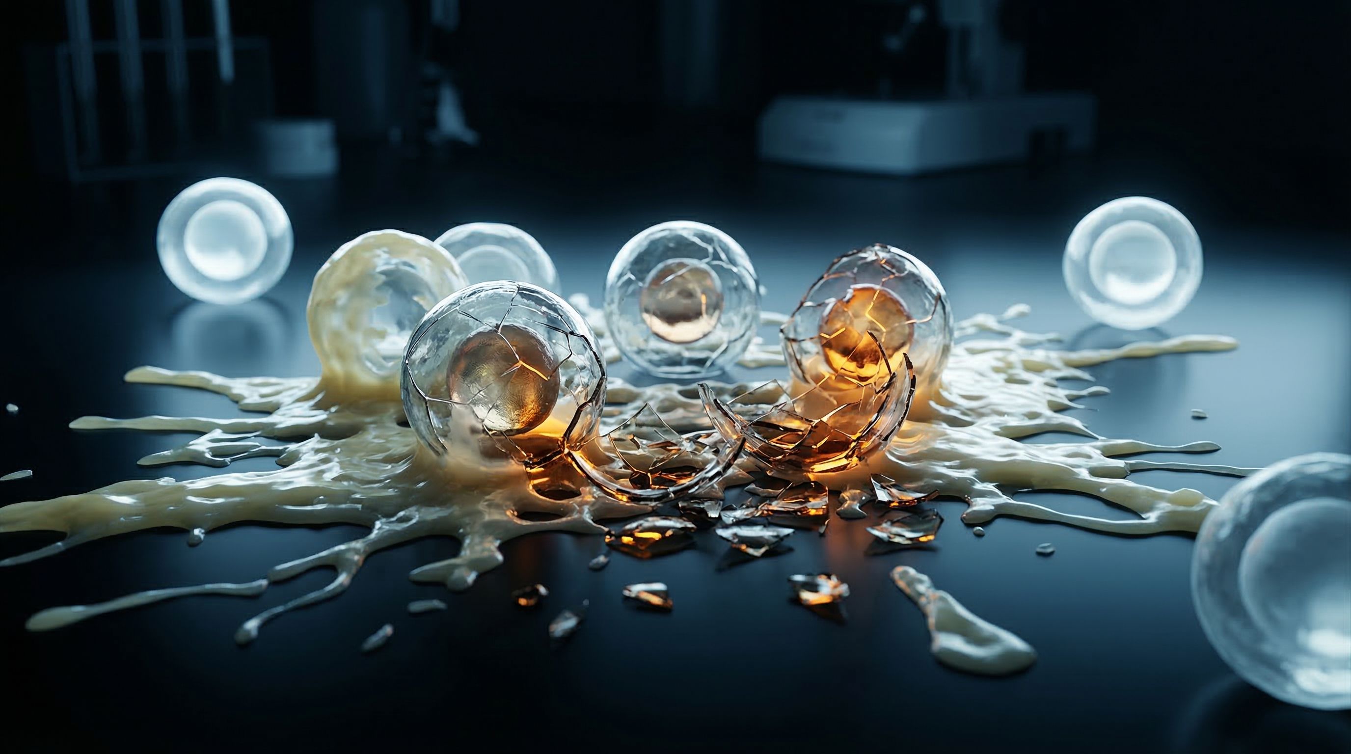
Getting useful information out of archived brain tissue is harder than it sounds. The formalin that preserved the tissue also damaged the RNA inside each cell, fragmenting it into shorter and shorter pieces over time. A block archived five years ago has better-quality genetic material than one archived twenty years ago. The older the case, the more degraded the evidence.
Then there is myelin, the fatty insulation coating nerve fibers. Brain tissue is loaded with it. When researchers try to break apart preserved tissue to access individual cells, myelin turns into a waxy debris that clogs equipment, contaminates samples, and makes everything harder.
And there is a third problem, one that rarely gets discussed outside the lab. The methods currently used to extract cells from these archived blocks are rough. They destroy a large fraction of the material. The most fragile cells, the neurons that researchers most want to study, are often the first casualties. What survives the extraction tends to be the tougher immune cells, leaving researchers with a skewed picture of what was actually in the tissue.
One shot at the evidence
Biobanks typically allocate a limited number of sections per researcher. A postmortem brain is not a renewable resource. If the extraction method destroys most of the material or returns biased results, there may not be enough tissue left for a second attempt. The evidence is decades old. You get one shot to crack the case.
Cold cases are being reopened
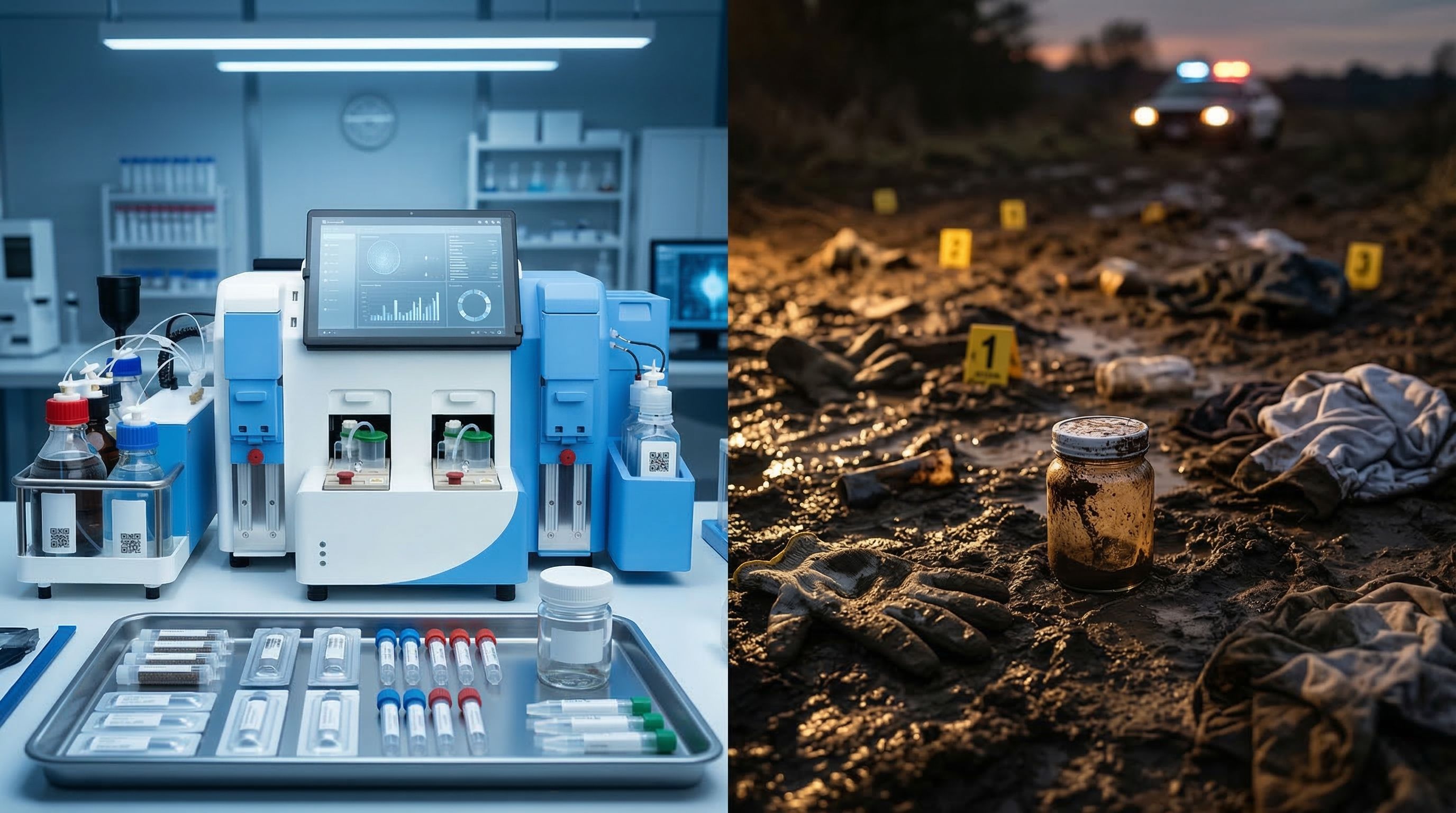
New sequencing technologies are changing the equation. Methods designed specifically for degraded tissue can now read the fragmented genetic material in archived samples. Spatial platforms can map gene activity across a tissue section without disrupting its architecture. These advances mean the evidence locked in paraffin is no longer inaccessible.
But technology is only half the answer. The other half is preparation, getting the cells or their nuclei out of the paraffin cleanly, consistently, and without destroying the very populations that hold the most information. That preparation step, the bridge between an archived block and usable data, is where most cold cases still stall.
In hospitals and research institutions around the world, the evidence sits waiting. Millions of brain tissue blocks, decades of clinical histories, thousands of patients whose donations have not yet delivered on their promise.
The cases are cold. But they are not closed.
In Part 2 of Cold Case, we examine why current methods for extracting evidence from these archived samples destroy so much of what they are trying to find, and what that costs neuroscience research.