FFPE Nuclei Extraction for Neurodegenerative Disease Research
The tissue that tells a twenty-year story
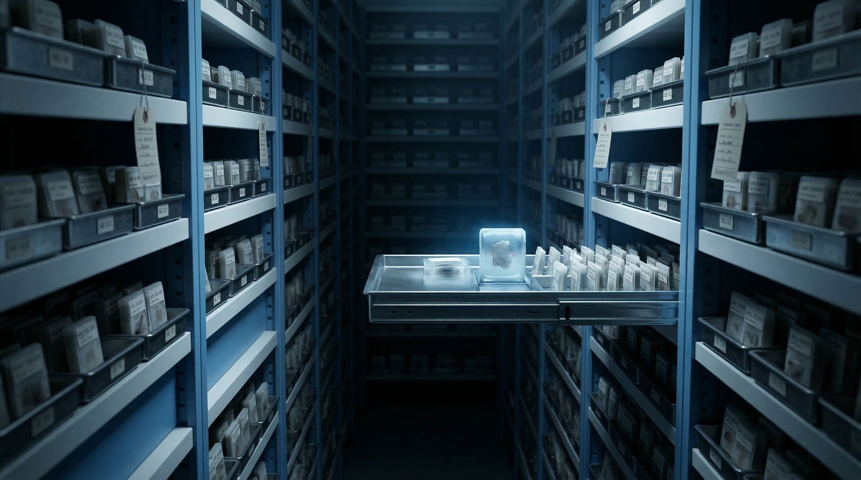
Somewhere in a brain bank freezer, there is an FFPE block from a patient enrolled in an Alzheimer's cohort in 2004. That patient was followed through annual cognitive assessments, amyloid PET imaging, CSF biomarker draws, and eventually autopsy. The block contains hippocampal tissue fixed at Braak stage V, and the clinical annotations attached to it represent two decades of longitudinal data that no prospective study could recreate.
Across the hall, another block holds substantia nigra from a Parkinson's patient whose dopaminergic neuron loss was documented over fifteen years of clinical visits. In a different building, a freezer holds frontal cortex sections from an ALS patient whose motor neuron degeneration progressed through three years of clinic records.
These blocks are not just tissue. They are the physical substrate of irreplaceable clinical narratives. And the method used to extract nuclei from them determines whether those narratives contribute to our understanding of neurodegeneration or become another set of failed preps in a lab notebook.
This guide covers specific strategies for processing FFPE brain tissue from neurodegenerative disease archives on the Singulator 200+, with attention to the challenges that make this work distinct from general FFPE processing: disease staging across Braak and other classification systems, longitudinal cohort comparisons spanning years of processing, cell-type vulnerability patterns where the neurons dying from disease are the same ones killed by harsh extraction, and the multi-site consortium reproducibility that programs like ADNI and AMP-AD require.
TL;DR -- Neurodegenerative disease FFPE essentials
- Disease-stage comparisons (Braak I vs. VI, early vs. late Parkinson's) require identical processing -- the S200+ delivers 1.0M/1.0M replicate consistency
- Fragile neuronal nuclei lost to manual processing are the same populations carrying disease signatures
- Blocks archived 10, 20, or 30+ years still yield usable nuclei -- probe-based sequencing handles degraded RNA
- Multi-site consortia (ADNI, ROSMAP, AMP-AD) need operator-independent results that manual methods cannot guarantee
- The two-cartridge workflow processes inputs as small as a single section from a brain bank allocation
Extract the biology that neurodegeneration is trying to teach you
Five deep-dive topics covering the specific challenges of working with FFPE brain tissue from neurodegenerative disease archives, from disease staging through multi-site consortium reproducibility.
Process tissue across disease stages Process FFPE brain tissue across neurodegenerative disease stages
Neurodegenerative disease research often requires comparing tissue from different disease stages within the same cohort. An Alzheimer's study may need hippocampal sections from Braak stage I (preclinical) through Braak stage VI (severe). A Parkinson's study may compare substantia nigra from patients with early motor symptoms to those with advanced disease. The scientific question is straightforward: which cell types change, and how, as disease progresses? But the sample prep cannot be allowed to introduce its own variability on top of the biological differences.
Why disease-stage comparisons are uniquely demanding
When you compare a Braak I section to a Braak VI section, any difference in the data should reflect disease biology -- not processing artifacts. Manual methods introduce variability at every step. If the Braak I section was processed by one technician on a Tuesday and the Braak VI section by a different technician three months later, the differences in deparaffinization timing, trituration force, and enzymatic incubation will create batch effects that overlap with the disease signal you are trying to measure. Computational correction can reduce these effects, but it cannot eliminate confounding that is systematically correlated with disease stage.
If your Braak I samples were all processed in month one and your Braak VI samples in month six, any processing-related batch effect is perfectly correlated with disease stage. No amount of batch correction can separate a genuine disease signature from a systematic processing artifact when the two variables are confounded. The only solution is to eliminate the processing variability at the source.
Staging systems and tissue selection
Different neurodegenerative diseases use different staging systems, and each system specifies distinct brain regions. Alzheimer's disease uses Braak staging for neurofibrillary tangles: entorhinal cortex in early stages, spreading to hippocampus and neocortex. Parkinson's uses Braak staging for alpha-synuclein pathology, progressing from brainstem to limbic to neocortical regions. Lewy body dementia overlaps with both. ALS has its own staging based on TDP-43 spread patterns. The brain region you section depends on the stage you are studying and the pathology you are tracking.
When designing a disease-stage comparison study, select brain regions where the pathology is expected to be present at that stage, not regions where you hope it might be. Processing hippocampus from a Braak I patient looking for hippocampal tangles will likely show a normal transcriptomic profile -- the tangles have not reached the hippocampus yet. Work backward from the staging system to select the appropriate tissue.
The FFPE two-cartridge workflow for disease staging studies
The Singulator 200+ processes each section through the same automated sequence: GREEN FFPE cartridge for deparaffinization, then YELLOW NIC+ cartridge for nuclei isolation. S200+ Only Every section receives identical mechanical force, identical enzymatic conditions, and identical timing. Whether you process the Braak I section today and the Braak VI section next month, the instrument removes the operator as a variable. Replicates yield 1.0 million and 1.0 million nuclei -- not the 1.5 million and 0.4 million variability seen with semi-automated methods.
Preserve the neurons disease is killing Preserve the vulnerable neuronal populations that disease targets
The central irony of neurodegenerative disease research with FFPE tissue is that the neurons most relevant to your study are the ones most likely to be destroyed by your sample prep. Alzheimer's targets hippocampal pyramidal neurons. Parkinson's targets dopaminergic neurons in the substantia nigra. ALS attacks motor neurons in the spinal cord and motor cortex. These are large, morphologically complex cells with extensive processes and fragile nuclei -- and manual trituration kills them preferentially.
Cell-type vulnerability maps to processing vulnerability
Neuronal nuclei are larger than glial or microglial nuclei. Hippocampal pyramidal neurons have nuclei measuring 10 to 15 micrometers in diameter, roughly twice the size of microglial nuclei. Larger nuclei are more susceptible to shear forces during mechanical dissociation. The vigorous pipetting required to break apart cross-linked FFPE tissue applies forces that exceed the structural integrity of these large neuronal nuclei, while smaller, denser microglial and oligodendrocyte nuclei survive. The result in your snRNA-seq data: microglial clusters dominate, neuronal clusters shrink, and the very cell populations undergoing disease-driven transcriptomic changes are underrepresented or absent.
The Singulator 200+ applies controlled, calibrated mechanical and enzymatic processing within sealed cartridges. The force is sufficient to dissociate cross-linked FFPE tissue but gentle enough to preserve large neuronal nuclei that manual trituration destroys. In comparable FFPE tissue studies, the S200+ enriched for fragile attached cell types that semi-automated methods lost, with semi-automated output skewing toward immune cells.
What biased cell-type recovery means for disease studies
If your Alzheimer's hippocampal dataset shows abundant microglia but few excitatory neurons, you cannot distinguish whether the neurons are absent because the disease destroyed them or because the sample prep did. This ambiguity is fatal for disease-stage comparisons, where the entire point is to track neuronal loss and transcriptomic change across progression. A prep method that consistently loses neurons creates a floor effect -- every stage looks like late-stage disease because the neurons are gone from the data regardless of what happened in the tissue.
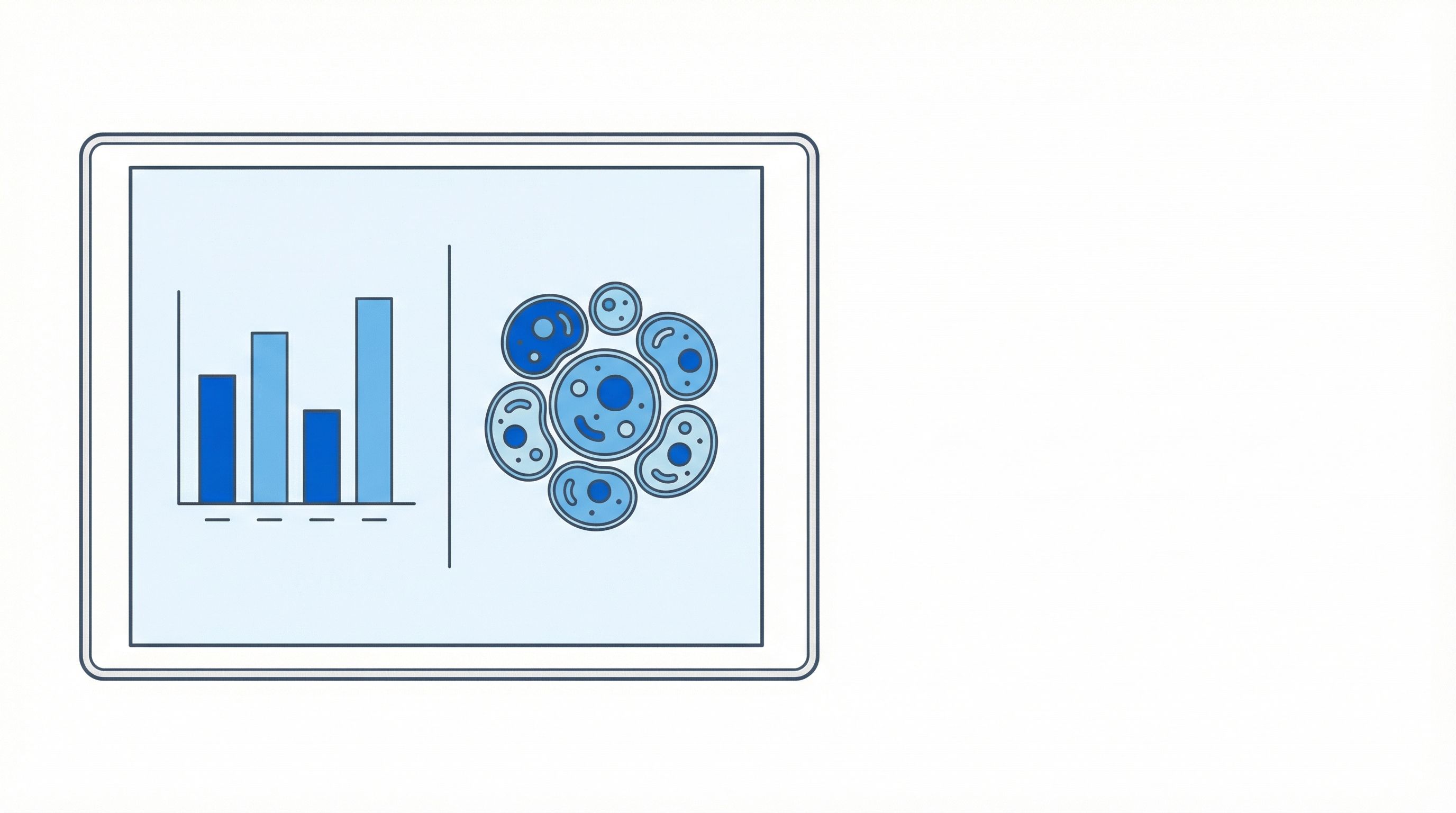
Compare your snRNA-seq cell-type proportions against published spatial transcriptomics data from the same brain region. Spatial methods analyze tissue in situ without dissociation, so they provide an unbiased reference for expected cell-type ratios. If your sequencing data shows far fewer neurons than the spatial reference, the extraction method is the likely bottleneck.
A dataset showing 80 percent microglia from hippocampal tissue may reflect genuine neuroinflammation -- or it may reflect a prep method that destroyed most neuronal nuclei. Without a reference for expected cell-type proportions, you cannot tell the difference. Use spatial data or published histological counts as your benchmark before drawing biological conclusions from cell-type ratios.
Compare longitudinal cohort tissue Compare tissue processed months or years apart within the same cohort
Longitudinal neurodegenerative disease studies can span decades. The Religious Orders Study and Rush Memory and Aging Project (ROSMAP) has been collecting postmortem brain tissue since 1994. The Alzheimer's Disease Neuroimaging Initiative (ADNI) has followed patients through multiple waves of data collection. When these cohorts reach autopsy, the tissue arrives at the lab in waves -- not all at once. The first brains may be processed years before the last ones.
The batch effect problem in long-running studies
Manual FFPE processing introduces variability at every step: deparaffinization timing, rehydration duration, trituration pressure, enzymatic incubation temperature. If your lab processes the first wave of ROSMAP tissue in 2024 with one technician and the second wave in 2027 with a different technician, the processing variability between waves may be larger than the biological differences between patients. Personnel change. People graduate, take new jobs, go on parental leave. The technician who developed excellent manual technique over two years is replaced by someone learning the protocol from a written SOP that describes "vigorous pipetting" without quantifying what vigorous means.
A longitudinal Alzheimer's study may process tissue from the first autopsy in year one and the last autopsy in year ten. Manual protocols cannot guarantee consistent results across that span. The S200+ delivers 1.0M/1.0M replicate consistency whether the samples are processed on the same day or years apart, because the instrument applies identical mechanical force and enzymatic conditions regardless of when the button is pressed.
What consistency actually means for your analysis
The Singulator 200+ reduces the processing workflow to 4 pipetting steps and less than 5 minutes of hands-on time. Compare that to the 28 pipetting steps and 25 minutes of hands-on time required by semi-automated methods. With fewer manual steps, there are fewer opportunities for human variability to enter the data. The 81 percent reduction in hands-on time is not about saving a technician twenty minutes -- it is about eliminating twenty minutes of potential batch effects from every sample in a multi-year cohort.
Archival block quality across cohort timepoints
A long-running cohort means that the first blocks in the study may be significantly older than the last ones. A brain autopsied and fixed in 2005 has been sitting in paraffin for twenty years by the time you process it in 2025. A brain from 2023 has been archived for only two years. RNA quality (measured by DV200) typically declines with block age, but nuclei integrity is more resilient. The S200+ recovers intact nuclei from blocks of varying age. For RNA quality, run a DV200 check on a thin test section before committing your limited curls.
| Block age | Expected DV200 | Recommended platform |
|---|---|---|
| 0-5 years | >50% | 10x Flex (strong data quality) |
| 5-15 years | 30-50% | 10x Flex (usable, reduced genes per cell) |
| 15+ years | <30% | Spatial (Xenium) or targeted panels |
If your cohort spans blocks from 2000 to 2024, do not assume all blocks will perform equally. Group your samples by approximate block age and run a pilot DV200 from each age group before committing precious sections. This prevents investing a rare Braak VI section from 2002 into a Flex run when the RNA quality may be better suited for a spatial approach.
Standardize across consortium sites Standardize processing across multi-site neurodegenerative disease consortia
Programs like ADNI, AMP-AD, the BRAIN Initiative Cell Census Network, and the Human Cell Atlas Brain Initiative distribute tissue and data across multiple institutions. The scientific value of these programs depends on comparability: data from Site A must be analyzable alongside data from Site B without site being a confounding variable. Manual FFPE processing makes this nearly impossible, because every site has its own technicians, its own interpretation of "vigorous trituration," and its own version of deparaffinization timing.
Why multi-site studies break with manual processing
Consider a four-site Parkinson's consortium where each site receives substantia nigra sections from the same patient cohort. With manual protocols, Site A uses 20-minute deparaffinization, Site B uses overnight because their technician starts the protocol at 4 PM and processes the next morning. Site C has a postdoc who uses gentle trituration; Site D has a technician who pipettes forcefully. The resulting nuclei preparations differ in yield, cell-type composition, and debris levels -- not because the tissue differed, but because the people differed. When these datasets are merged, site-specific batch effects can mask or mimic disease biology.
The Singulator 200+ replaces operator-dependent manual steps with a fixed, automated workflow. The GREEN FFPE cartridge runs deparaffinization on-instrument with a proprietary safe solvent -- no fume hood required, which means any site can participate regardless of lab setup. The YELLOW NIC+ cartridge runs nuclei isolation with pre-installed protocols. Four pipetting steps. Less than 5 minutes hands-on. What remains for the human to vary is almost nothing.
Practical deployment across sites
For a consortium deploying the S200+ across multiple institutions, the workflow is the same at every site: load tissue into GREEN cartridge, run deparaffinization, transfer to YELLOW NIC+ cartridge, run nuclei isolation, collect output. No site-specific protocol adaptations. No local deparaffinization timing preferences. No variability in trituration force. The instrument is the protocol, and the protocol is identical everywhere.
The elimination of toxic solvents (no xylene, no CitriSolv) removes a practical barrier to multi-site deployment. Sites without fume hoods or chemical waste disposal infrastructure can still run the FFPE workflow. This matters for smaller academic institutions or satellite facilities that might otherwise be excluded from a consortium because of facility limitations.
Share the replicate data with consortium leadership: S200+ replicates yield 1.0M and 1.0M nuclei versus 1.5M and 0.4M with semi-automated methods. That 3.75-fold improvement in consistency translates directly to reduced batch effects when merging data across sites. For an NIH-funded consortium, processing standardization is often an explicit review criterion.
Choose the right analytical platform Choose the right analytical platform for your neurodegenerative disease question
The analytical platform you choose should match your scientific question, not the other way around. A study tracking transcriptomic changes in specific neuronal subtypes across Braak stages needs sequencing depth. A study mapping where pathology-associated cell types sit within brain architecture needs spatial resolution. Many studies need both. The Singulator 200+ produces platform-agnostic nuclei, which means you can design your analytical strategy based on the biology rather than the sample prep.
10x Genomics Flex for degraded archival tissue
The Flex assay is the default choice for snRNA-seq from neurodegenerative disease FFPE tissue. Its probe pairs have a compact 50-nucleotide footprint, bypassing the need for intact poly-A tails -- which is relevant for archival blocks where RNA degradation is expected. FFPE tissue from PDAC studies processed on the S200+ yielded 1,844 to 2,245 UMI counts per nucleus on Flex, demonstrating that the nuclei quality supports meaningful transcriptomic data even from fixed tissue. A single brain section processed on the S200+ typically yields over 1 million nuclei -- far exceeding the 10,000 to 20,000 needed for a Flex loading.
10x Flex supports sample multiplexing, which allows you to pool nuclei from multiple disease stages onto a single chip. Process each Braak stage section individually on the S200+, then pool the nuclei at known ratios before Flex loading. This approach reduces per-sample sequencing cost while enabling direct comparison of disease stages within the same sequencing run, minimizing technical batch effects between stages.
Spatial transcriptomics for architectural context
Neurodegenerative diseases alter brain architecture -- amyloid plaques in Alzheimer's, Lewy bodies in Parkinson's, motor neuron loss patterns in ALS. Spatial platforms like 10x Xenium map these patterns in situ. Memorial Sloan Kettering's Dana Pe'er lab used S200+ nuclei alongside Xenium spatial analysis for brain tissue studies. For neurodegenerative disease research, consider sectioning adjacent sections from the same block: one for Xenium spatial analysis and one for S200+ nuclei extraction and snRNA-seq. The spatial data provides the map; the snRNA-seq provides the depth to annotate every cell type on that map.
PERFF-seq for rare disease-specific populations
Some neurodegenerative disease questions target rare populations: disease-specific neuronal subtypes undergoing selective degeneration, infiltrating immune cells at disease boundaries, or reactive astrocyte subsets around plaques. PERFF-seq, validated by Stanford and Memorial Sloan Kettering, captures rare cells from FFPE tissue at depths that standard snRNA-seq cannot match. S200+ nuclei are validated for this workflow. When your question hinges on a cell population that constitutes less than 1 percent of the tissue, PERFF-seq maximizes the information you extract per nucleus.
For irreplaceable neurodegenerative disease tissue, commit to a platform before cutting the block. Each platform has different input requirements and different tolerance for debris and RNA quality. Discovering after extraction that your nuclei are insufficient for the intended platform means wasting tissue you cannot replace. Plan the analysis, then work backward to the section.