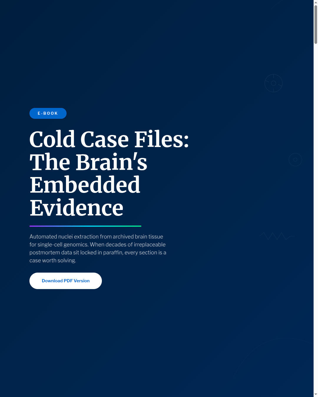
Cold Case - Part 2:
Contaminated Evidence
Cold Case: When Brain Tissue Holds the Evidence - Part 2 of 5
(Prefer to listen to the full story? Click play to hear Episode 2 of our accompanying audio version.)
The Precision Point - Cold Case - Episode 2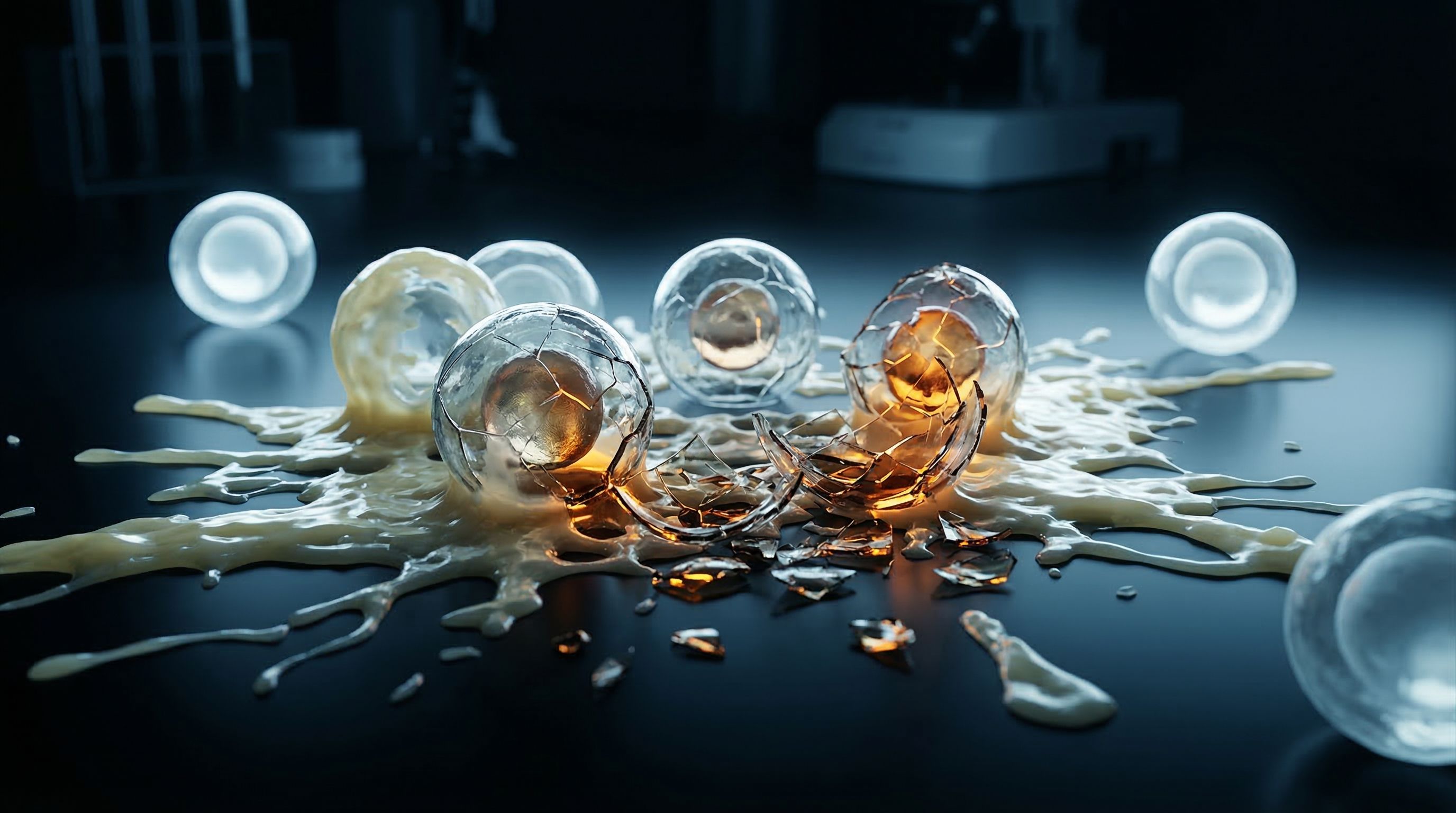
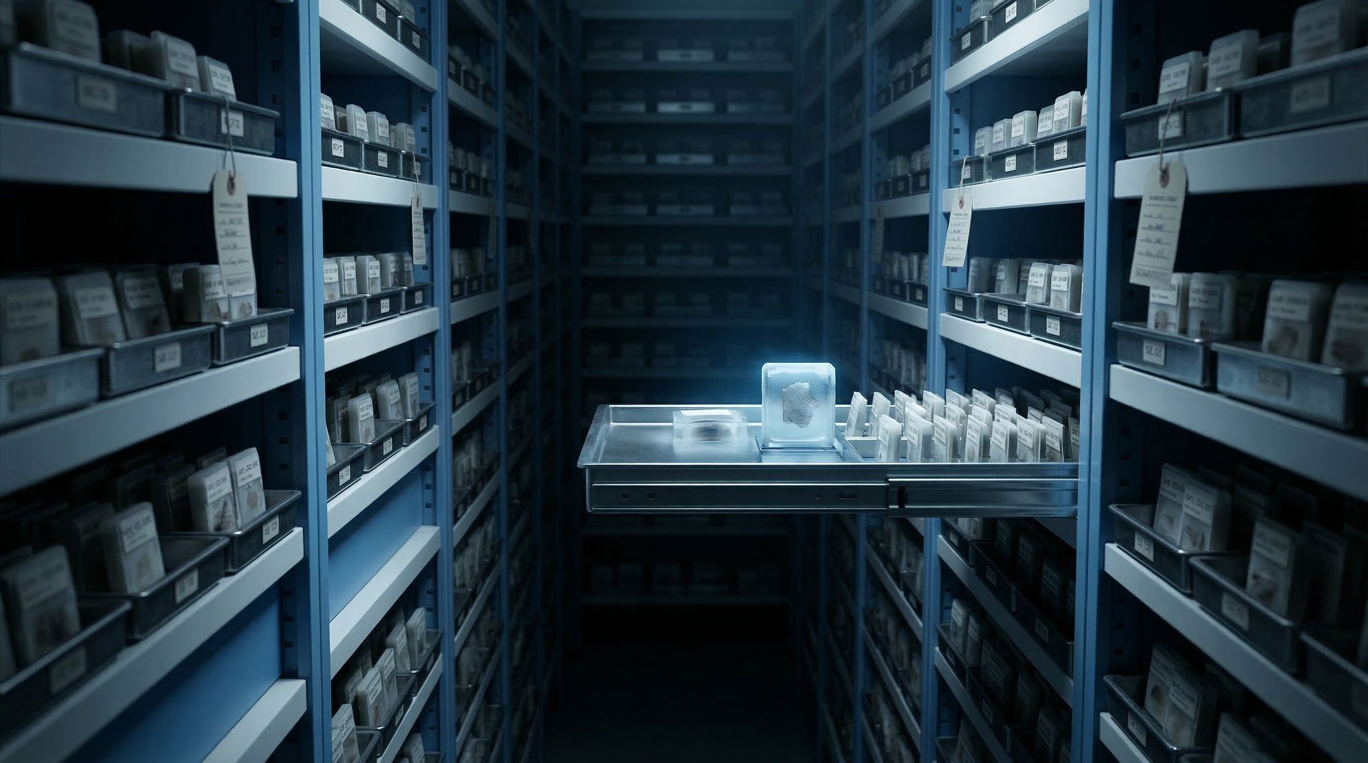
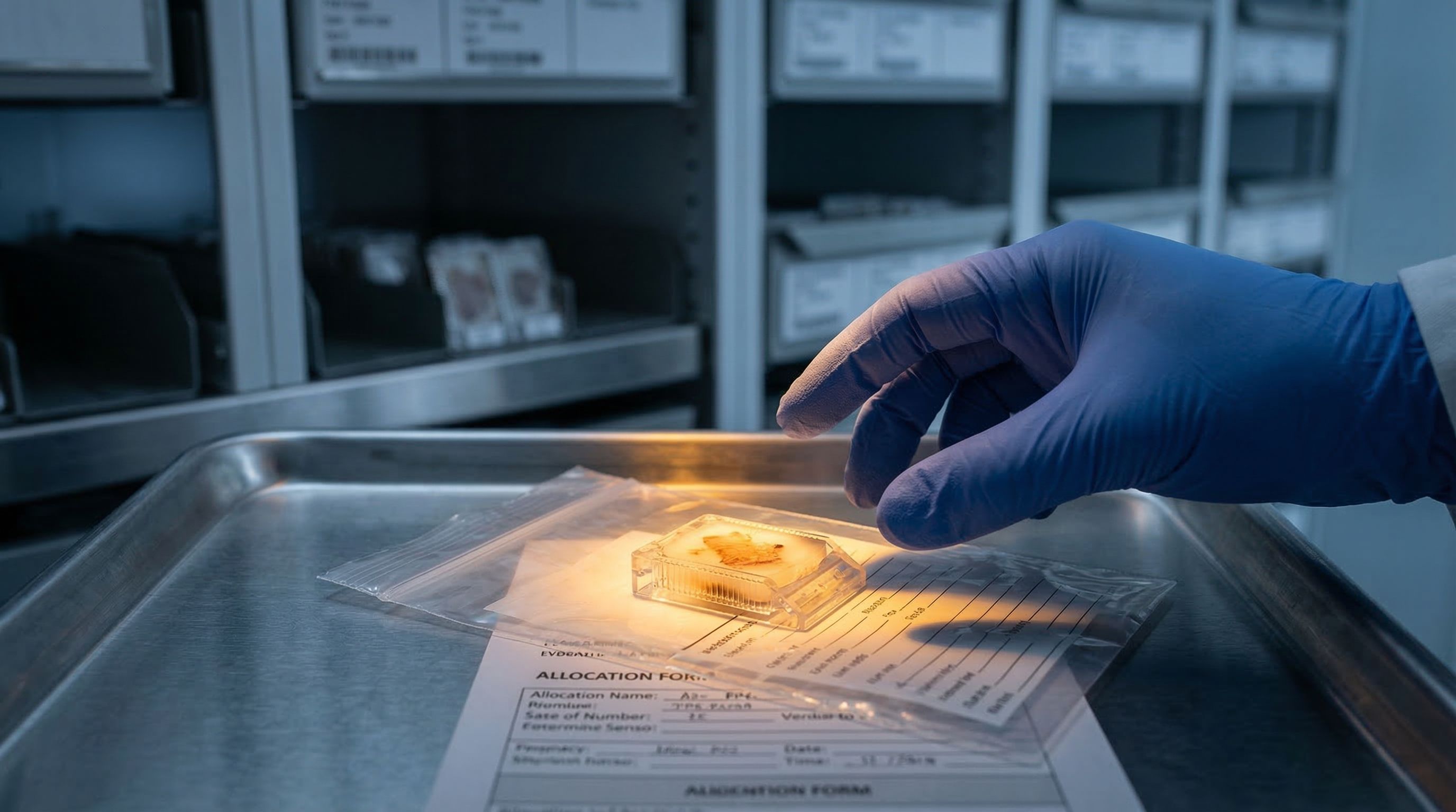
In Part 1 of Cold Case, we opened the evidence locker. Millions of brain tissue blocks sitting in paraffin archives, each one containing potential clues about Alzheimer's, Parkinson's, and dozens of other diseases. The evidence is there. The witnesses are preserved.
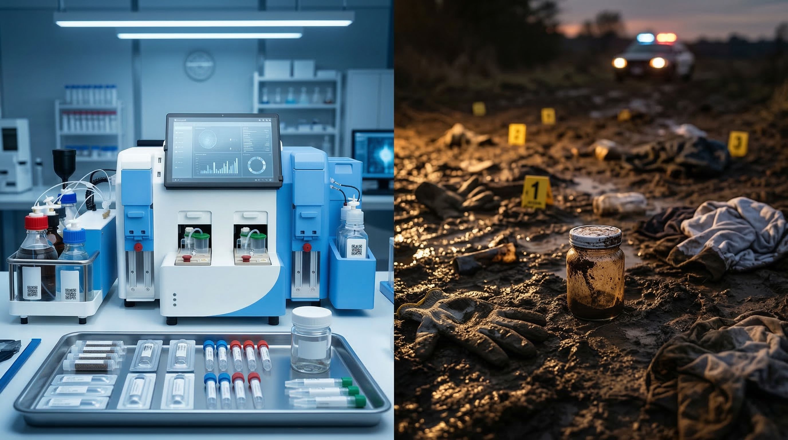
Now imagine a detective finally gets permission to reopen one of those cases. She walks into the crime scene, ready to collect evidence with modern forensic tools. But the first officer on the scene already trampled through it. Footprints over footprints. Key samples bagged and mixed together. Fragile trace evidence crushed under careless handling.
That is roughly what happens every time a researcher processes archived brain tissue using standard laboratory methods.
Trampling the crime scene
The standard approach to getting cells or nuclei out of archived brain tissue is manual dissociation. A researcher takes a section of preserved tissue, strips away the paraffin wax that has been protecting it, and then uses a combination of chemicals and physical force to break the tissue apart into individual cells or their nuclei.
The process takes three to five hours. It involves toxic solvents like xylene for removing the wax, a series of ethanol washes, enzymatic digestion, and repeated mechanical disruption, the scientific term for mashing tissue with a pestle or forcing it through a pipette tip over and over.
Each of these steps has a purpose. Each one also destroys evidence.
The damage report
Manual extraction methods lose 50 to 60 percent of the starting brain tissue during processing. Of the material that survives, only about a third is intact nuclei. The rest is cellular debris, partially destroyed cells, and fragments. To put it plainly: for every three pieces of evidence that go in, roughly one usable piece comes out.
Those numbers would be bad enough if the losses were random. They are not.
The witnesses who die first
Brain tissue contains many different cell types, and they do not all respond the same way to rough handling. Neurons are fragile. They have long, thin projections called axons and dendrites, complex internal structures, and membranes that rupture easily under mechanical stress. When you mash brain tissue with a pestle, neurons break first.
The cells that tend to survive manual processing are the tougher ones: immune cells like microglia and macrophages, which evolved to be mobile and resilient. In forensic terms, the witnesses who had the most to say about the crime have been destroyed, and the ones left standing are a biased sample of who was actually at the scene.
This is not a theoretical problem. When researchers compare cell-type profiles from manually processed brain tissue to what spatial methods reveal in intact sections, the manual results consistently underrepresent neurons and overrepresent immune populations. The detective's final report describes a scene that does not match what actually happened.
The grease on the evidence
Brain tissue has another property that makes processing especially messy. About half its dry weight is lipid, fat. Much of that comes from myelin, the insulating sheath that wraps nerve fibers. Myelin itself is roughly 80 percent lipid by dry weight. It is what makes the brain feel greasy if you have ever handled one.
When brain tissue is broken apart during dissociation, myelin disintegrates into a fine waxy debris that gets everywhere. It coats equipment. It clogs filters. It contaminates the nuclei suspension. Running a myelin-contaminated sample through a single-cell sequencing platform is like trying to dust for fingerprints with grease on your gloves. The data that comes back is noisy, incomplete, and mixed with background signal from the debris.
Why brain is harder than other tissues
Liver, kidney, tumor samples from other organs all present challenges for dissociation. But brain tissue combines three problems at once: extreme cellular fragility (neurons), massive lipid contamination (myelin), and the additional damage from formalin fixation that has been sealing the tissue shut for years or decades. Most extraction protocols were developed on less demanding tissues and adapted for brain. The adaptation does not work well.
The operator problem
Even if the method were gentle enough to preserve fragile cells and clean enough to manage myelin, there is a third problem. Manual processing depends on the person doing it.
How long did they soak the tissue in xylene? Did they check the deparaffinization at 20 minutes, or did they step away and come back at 45? How hard did they pipette during trituration? How many times? What temperature was the enzyme bath?
These variables change from operator to operator, from day to day, sometimes from morning to afternoon. Two researchers following the same published protocol on the same tissue type can get dramatically different results. One run might yield 1.5 million nuclei. The next, from the same block, might yield 400,000. That kind of variability would be unacceptable in any other forensic discipline. In neuroscience sample preparation, it is normal.
What contaminated evidence costs
The consequences accumulate. A researcher receives a single section from a 20-year Alzheimer's cohort. The tissue is irreplaceable. She processes it manually. Sixty percent of the material is lost. The surviving nuclei are skewed toward immune cells. The myelin debris that made it into the sequencing prep adds noise to the data. And because the results are operator-dependent, nobody can be sure whether the cell-type proportions in the final dataset reflect the actual tissue or just the processing method.
The case is not solved. The evidence is contaminated. And there is no second section to try again.
The chain of custody is broken
In forensic science, chain of custody means documenting every step of evidence handling to ensure integrity. In brain tissue processing, the chain breaks at the first step. Every manual intervention introduces variability. Every operator adds their own signature to the results. There is no standardized record of how much force was applied, how long each step took, or what the conditions were. The evidence changes depending on who handles it.
The problem is not that researchers are careless. The problem is that the tools available for this work were never designed for tissue this fragile, this contaminated with lipid debris, or this irreplaceable. Methods that work well enough on fresh tumor tissue or cultured cells fall apart when applied to decades-old, formalin-fixed brain.
Standardized, automated approaches to this problem do exist, and a growing number of neuroscience labs are adopting them. We will get to what those look like. But first, in Part 3, we confront the constraint that makes everything harder: you get one shot at the scene. One section. One chance. And the consequences of a failed prep are not just a bad dataset. They are a permanently lost window into disease.