Cold Case - Part 3:
One Shot at the Scene
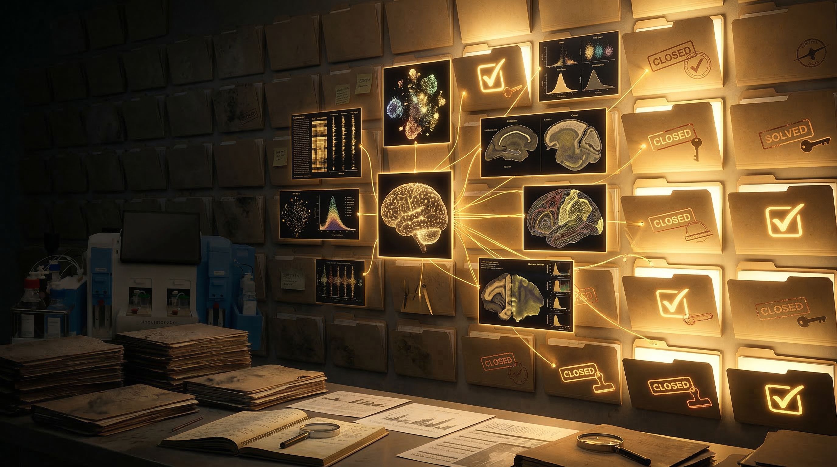
Cold Case: When Brain Tissue Holds the Evidence - Part 3 of 5
(Prefer to listen to the full story? Click play to hear Episode 3 of our accompanying audio version.)
The Precision Point - Cold Case - Episode 3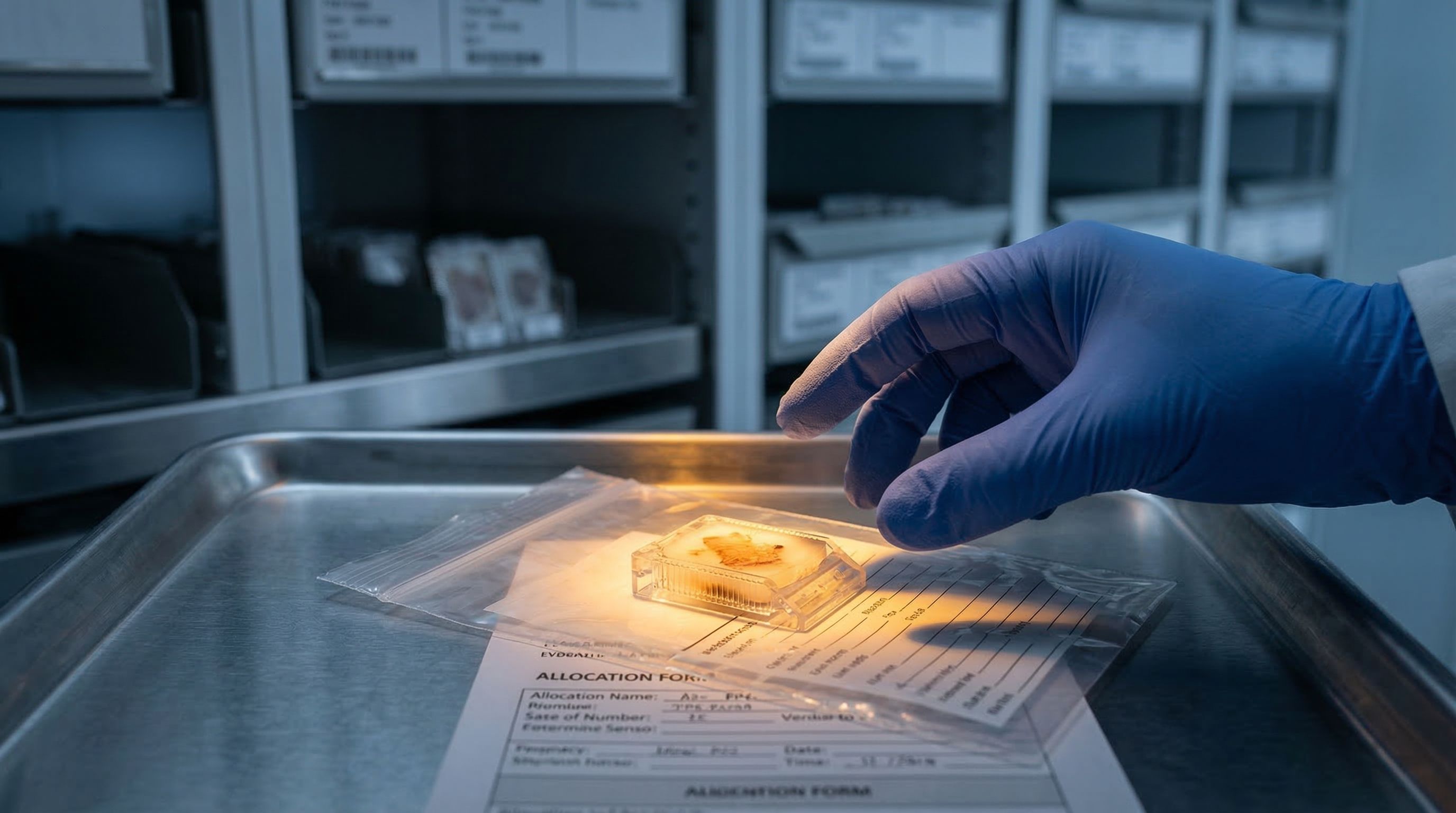
The email from the biobank coordinator is three sentences long. Your tissue request has been approved. You will receive four sections from a single FFPE block. There will be no additional allocations from this case.
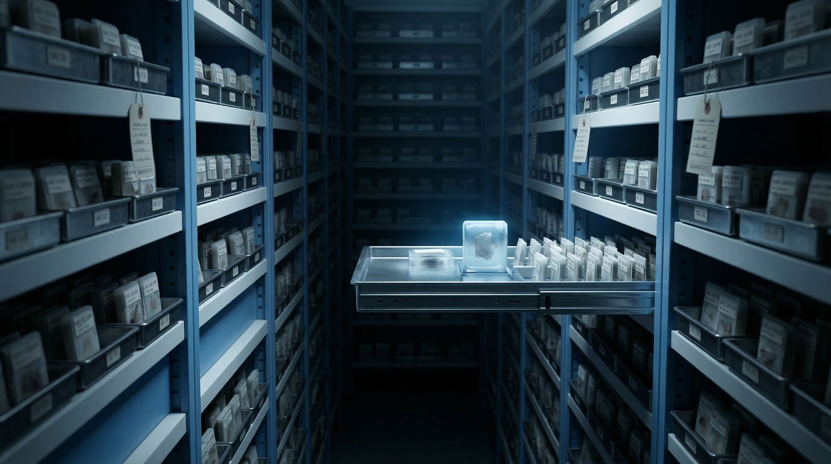
Four sections. From a patient who was followed for eighteen years through a progressive Alzheimer's diagnosis. Cognitive testing, brain imaging, detailed clinical notes at every stage. The patient died. The family consented to brain donation. The tissue was fixed, embedded, archived. And now it sits in front of you, cut into sections thinner than a credit card.
This is your crime scene. You do not get to visit it twice.
The allocation problem
Researchers who work with archival brain tissue know this pressure well. NIH-funded brain banks like the Harvard Brain Tissue Resource Center, the NIH NeuroBioBank, and the Mount Sinai Brain Bank operate under strict allocation policies. A typical request yields a handful of sections from each case. The tissue is finite. The demand is not.
For rare diagnoses, the constraint tightens further. Lewy body dementia, frontotemporal degeneration, progressive supranuclear palsy. Some of these conditions appear in fewer than a hundred cases across all major brain banks combined. A researcher studying one of these diseases might wait months for an allocation and receive sections from just a few patients.
Hospital pathology departments hold their own archives, but access follows similar rules. Tissue from a surgical resection, a tumor biopsy, an autopsy ordered by a neuropathologist. The blocks sit in storage, catalogued and numbered, but each section cut from a block is a section that no one else will ever use.
The forensic analogy holds here with uncomfortable precision. In a murder investigation, the detective gets one visit to the crime scene before it is cleaned and returned to the family. Everything the case will ever know must be collected in that visit. Every footprint, every fiber, every trace of evidence.
A researcher opening a package from a brain bank is standing at that threshold.
What happens when evidence is mishandled
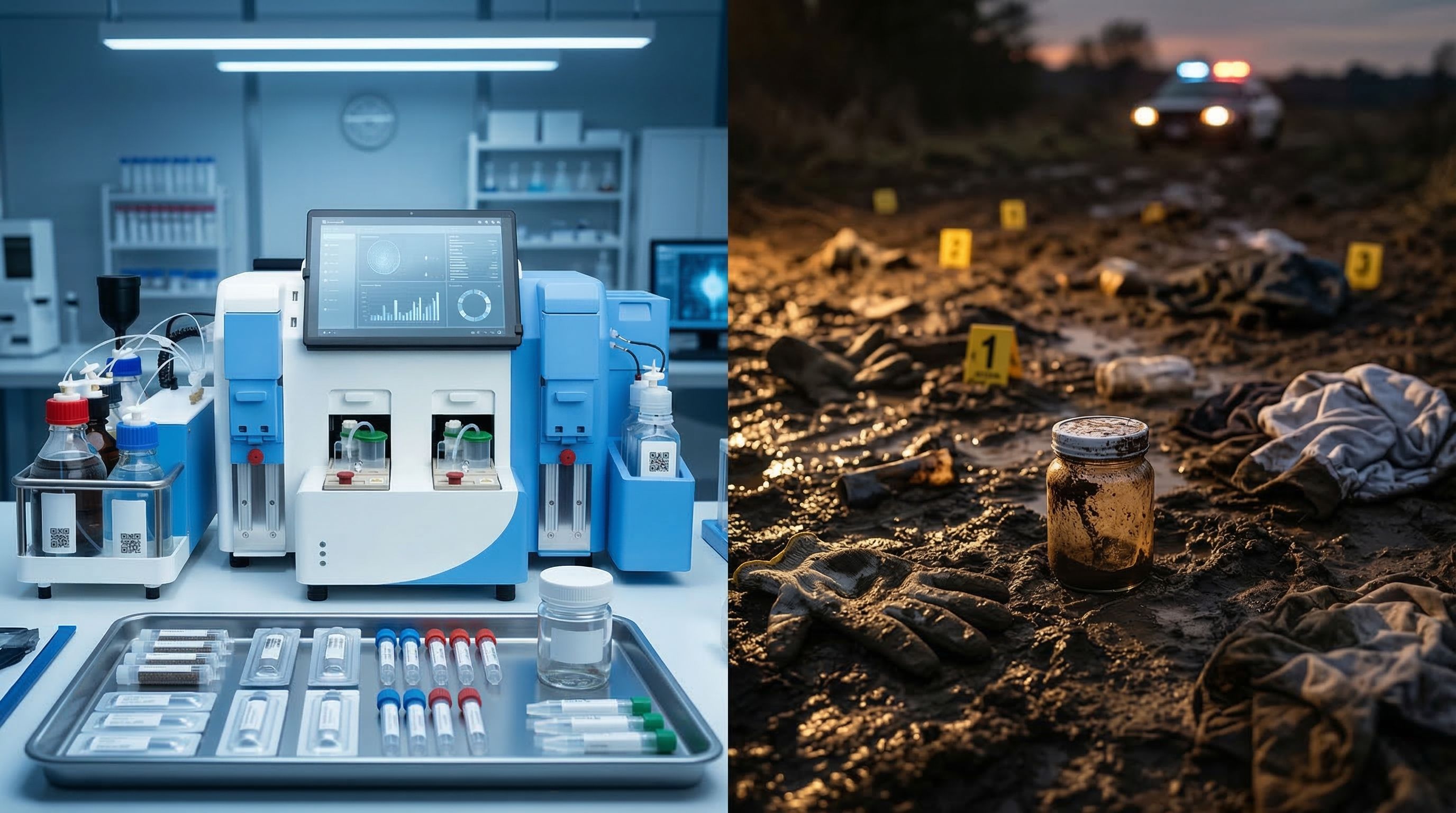
The standard approach to extracting nuclei from FFPE brain tissue involves manual processing. Deparaffinization with xylene or similar solvents. A graded ethanol rehydration series. Enzymatic digestion. Mechanical trituration with a pestle or by pipetting. Filtration. Each step performed by hand, each step introducing variability.
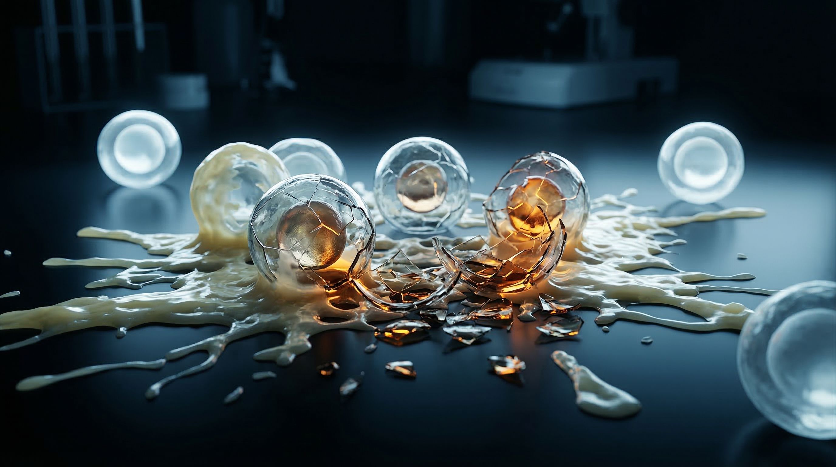
The numbers from this process are hard to read without wincing. Published protocols and field evaluations report that 50 to 60 percent of the starting tissue is lost during processing. Of what remains, only about a third are intact nuclei. The rest is a mixture of debris, cell ghosts, and partially digested material.
For brain tissue specifically, the losses are not random. The most fragile cells die first. Neuronal nuclei, the population that carries the most direct information about neurodegenerative disease, break under harsh mechanical force. What survives tends to be the hardier immune cell populations. The result is a dataset that over-represents microglia and under-represents the neurons and glia that researchers are most trying to understand.
The evidence you lose
When manual processing destroys 50 to 60 percent of a precious brain section, those lost cells are not a random sample. Fragile neuronal nuclei are disproportionately destroyed. The surviving population skews toward robust immune cells. The biological story that emerges from sequencing is distorted at the source, before a single read is generated.
Think about what that means for the Alzheimer's case sitting on your bench. You have four sections from an eighteen-year longitudinal study. If your processing method loses half the tissue and biases what remains, you are not just losing material. You are losing the specific cells whose transcriptomic data could explain why this patient's disease progressed the way it did.
You cannot go back to the biobank and ask for more. That crime scene is closed.
The twist: the method is the variable
Here is what makes Part 3 of a cold case investigation different from Parts 1 and 2. The first two parts establish what happened and who was harmed. Part 3 is where the investigation turns inward. What if the detectives themselves are compromising the evidence?
In neuroscience FFPE research, the parallel is uncomfortable but accurate. The biggest threat to your data is not the age of the tissue, or the quality of the formalin fixation, or even the fragmentation of the RNA. Those are real problems, but they are problems the field has tools to address. Probe-based sequencing handles fragmented RNA. Quality metrics like DV200 tell you what you are working with before you commit.
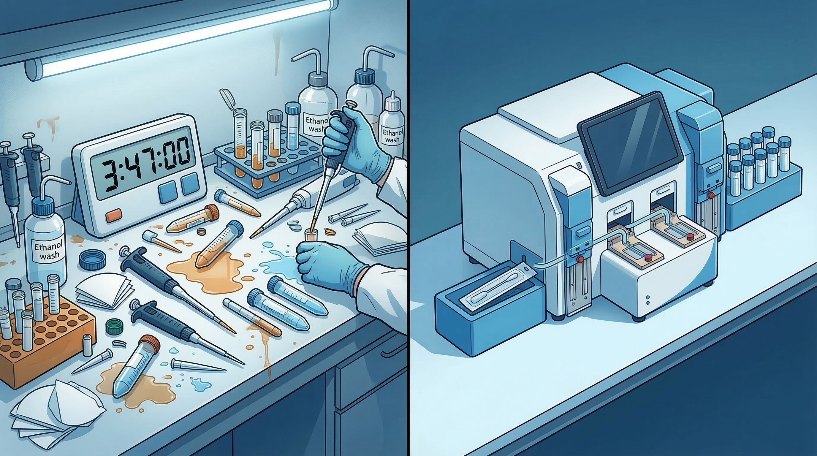
The biggest threat is the extraction step itself. Twenty-eight pipetting steps performed by hand. Three to five hours in a fume hood with toxic solvents. Deparaffinization timing that varies between operators. Trituration force that changes depending on who holds the pestle and how tired they are at hour four. A single protocol, performed by different people on different days, producing wildly different results.
One study comparing replicate samples found that the same protocol produced 1.5 million nuclei from one run and 400,000 from the next. A 3.75-fold difference. Not from different tissue. Not from different patients. From the same sample, processed the same way, by the same method.
The question changes
When your tissue is irreplaceable and your processing method loses half the material while biasing what survives, the question stops being "Can we extract nuclei from FFPE brain tissue?" and becomes "Can we afford to extract them the way we have been doing it?"
What a different approach looks like
The cold case analogy points toward its own resolution. When a police department realized that its detectives were contaminating crime scenes through inconsistent evidence collection, the solution was not to train the detectives harder. It was to build a forensic laboratory where the process itself became standardized. Same procedures, same conditions, same results, regardless of who ran the analysis.
The same logic applies to FFPE brain tissue processing. The variability does not come from the science. It comes from the twenty-eight manual steps that stand between an archived block and usable nuclei.
An automated approach that handles deparaffinization, enzymatic digestion, and nuclei isolation within a sealed, controlled system changes the math. Instead of twenty-eight pipetting steps, four. Instead of three to five hours of hands-on work in a fume hood, less than five minutes. Instead of operator-dependent results, instrument-controlled consistency.
The Singulator 200+ from Precision Cell Systems was designed for exactly this scenario. It automates the complete FFPE nuclei extraction workflow through a two-cartridge system. The first cartridge handles deparaffinization using a proprietary solvent that requires no fume hood. The second handles nuclei isolation. The operator loads the tissue, installs the cartridges, and walks away.
From a single 50-micrometer FFPE curl, the system consistently yields over one million nuclei. Replicates produce matching results: 1.0 million and 1.0 million, not 1.5 million and 0.4 million. The fragile neuronal populations that manual methods destroy are preserved, because the mechanical force is controlled by the instrument, not by a tired pair of hands at hour four of a fume hood protocol.
For the researcher holding four sections from an irreplaceable Alzheimer's case, the difference is not abstract. It is the difference between a dataset that reflects the actual biology of the tissue and one that reflects the limitations of the processing method.
One shot
The biobank coordinator's email does not care about your processing method. It tells you what you already know: the tissue is allocated, the allocation is final, and the science you produce from these sections is the only science anyone will ever produce from them.
The evidence is decades old. The patient's clinical history stretches back further. The family's decision to donate was made once.
How you process that tissue is the last decision left. It is also the one that determines whether the case stays cold or finally gives up its answers.
In Part 4 of Cold Case, we step inside the forensic lab. How automated processing preserves what manual methods destroy, and what the data looks like when the evidence is handled with the care it deserves.