Cold Case - Part 4:
The Forensic Lab
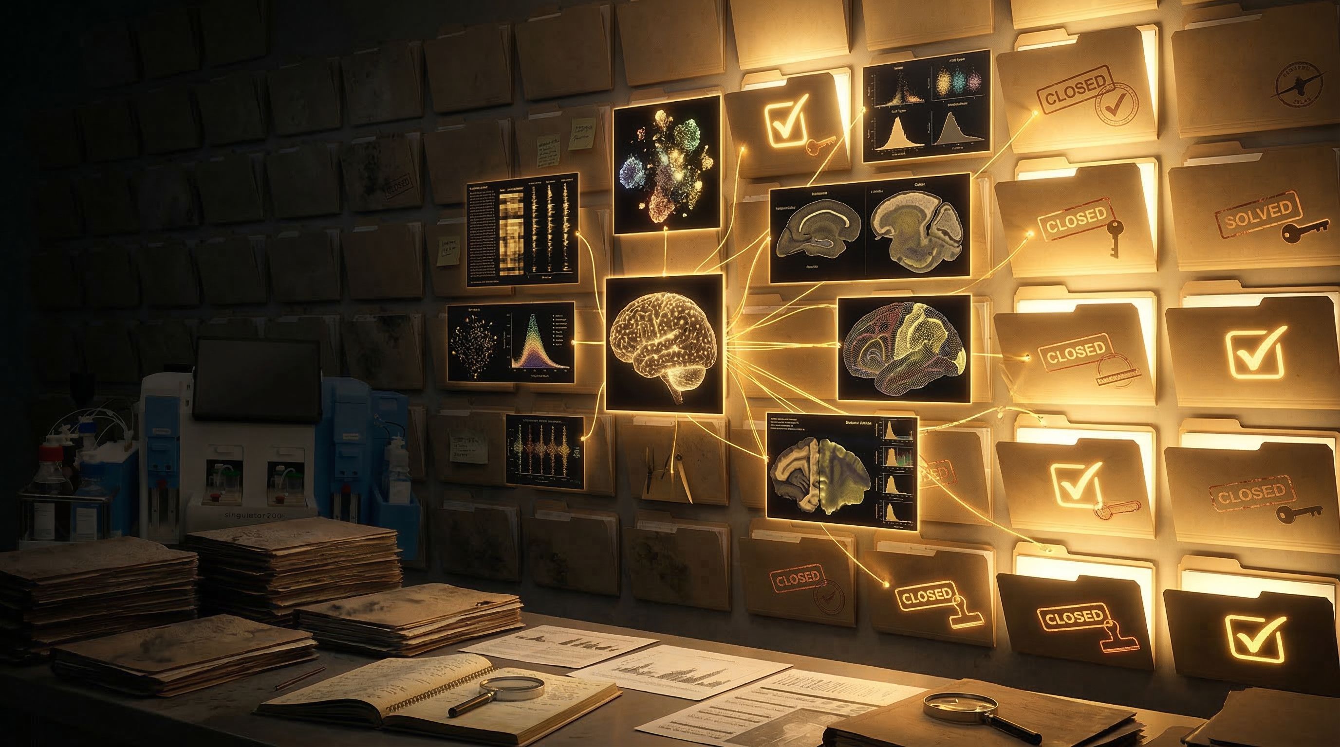
Cold Case: When Brain Tissue Holds the Evidence - Part 4 of 5
(Prefer to listen to the full story? Click play to hear Episode 4 of our accompanying audio version.)
The Precision Point - Cold Case - Episode 4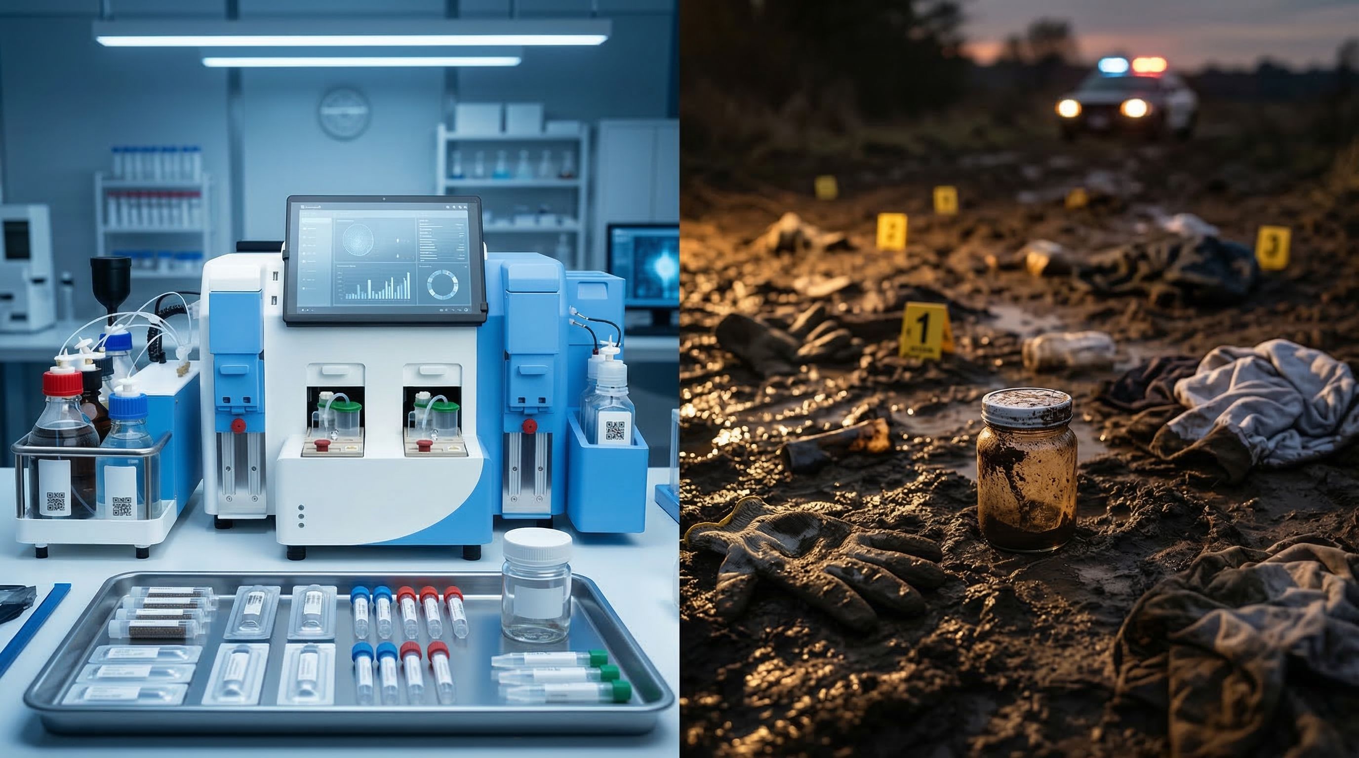
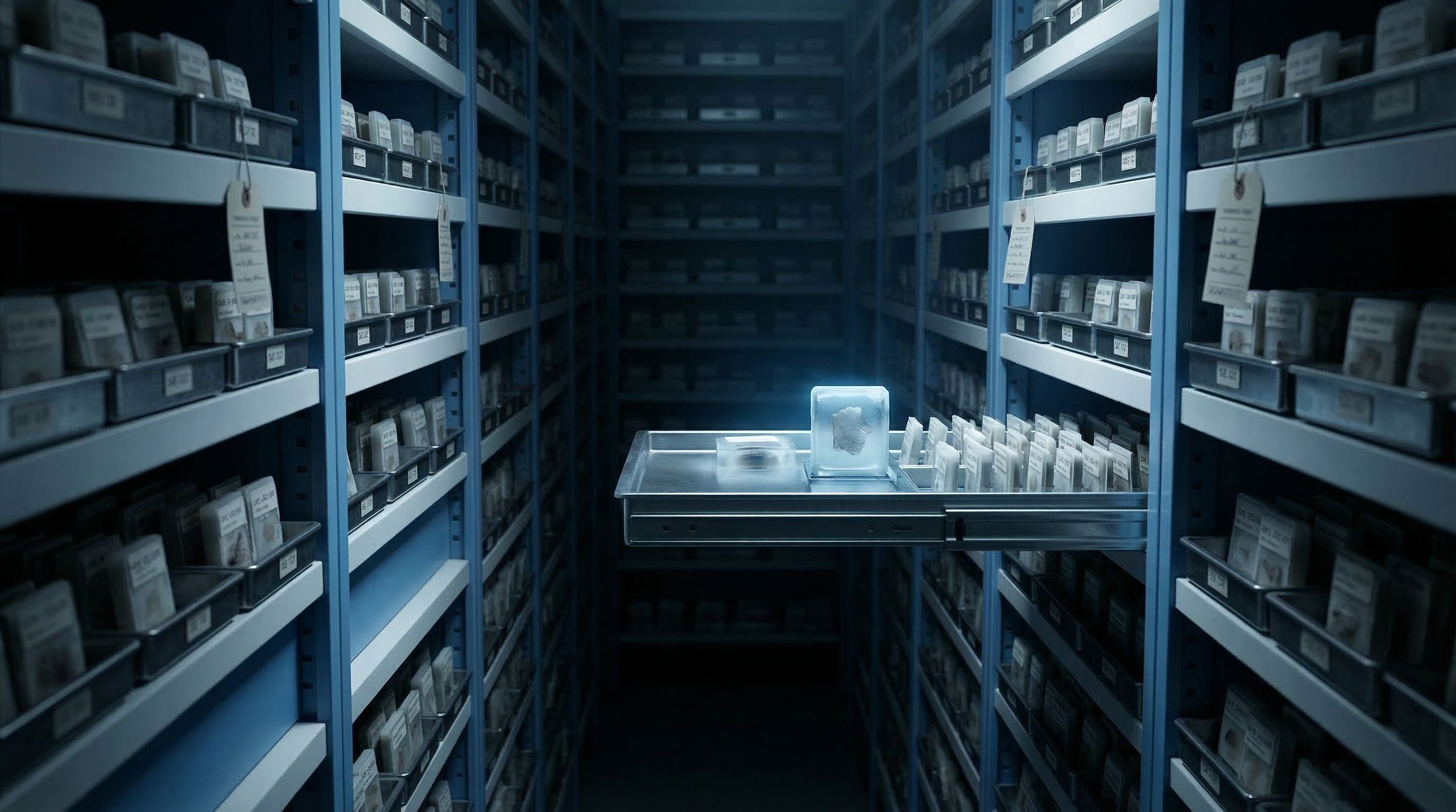
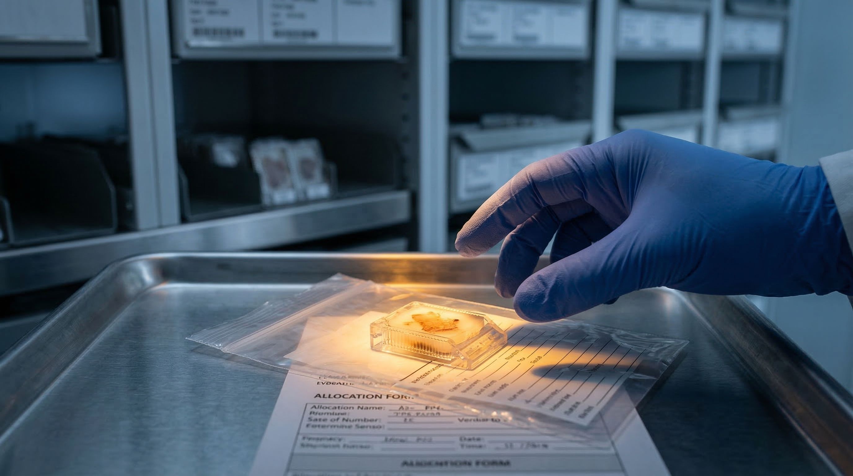
In criminal forensics, there is a moment when a case moves from the field to the lab. The muddy boot prints, the contaminated fibers, the partial fingerprints collected under rain, all of it gets transferred to a climate-controlled room where the rules change. The lighting is consistent. The instruments are calibrated. The technician running the analysis today will get the same result the technician running it next Tuesday would get.
That transfer, from uncontrolled fieldwork to controlled laboratory processing, is what separates modern forensic science from its predecessors. And for neuroscience FFPE research, that same transfer is now possible.
The problem with field processing
Consider what we described in Parts 2 and 3 of this series: a researcher standing at a bench, manually extracting nuclei from a postmortem brain section. The process takes three to five hours. It requires toxic solvents in a fume hood. There are 28 separate pipetting steps, each one an opportunity for variability. Trituration force depends on the researcher's wrist. Deparaffinization timing depends on whether someone checks the clock. Rehydration might sit overnight because the researcher went home.
This is field processing. It works, but it works the way crime scene collection worked before forensic labs existed: results depend on who showed up, how careful they were, and what the conditions were like that day.
For irreplaceable postmortem brain tissue, the stakes of that variability are permanent. A section from a twenty-year Alzheimer's cohort, processed by a departing postdoc on a Friday afternoon, produces different results than the same tissue type processed by the lab manager on a Tuesday morning. The biology has not changed. The operator has.
What a forensic lab looks like
The Singulator 200+ from Precision Cell Systems is the forensic lab for FFPE brain tissue processing. The comparison is not a marketing stretch. Consider what a modern forensic laboratory actually provides: standardized procedures that do not depend on the skill of individual technicians, sealed processing environments that prevent contamination, documented chain of custody at every step, and results that hold up to scrutiny because they were produced under controlled conditions.
The Singulator 200+ does the same thing for nuclei extraction. The entire process runs inside sealed cartridges. Two cartridges, used in sequence: a GREEN cartridge handles deparaffinization, the step that, in manual processing, is the single largest source of variability. Then a YELLOW NIC+ cartridge handles nuclei isolation and cleanup. The tissue goes in. Clean, sequencing-ready nuclei come out.
The two-cartridge chain of custody
GREEN cartridge: deparaffinization using a proprietary safe solvent, no xylene, no fume hood, no timing drift. YELLOW NIC+ cartridge: enzymatic digestion and nuclei isolation with built-in filtration that removes the myelin debris that plagues brain tissue preparations. The cartridges control what human hands cannot: identical conditions, identical timing, every run.
Operator-independent results
The most striking feature of the Singulator 200+ workflow is how little the operator matters. That sentence would be insulting in most laboratory contexts. Here, it is the point.
A manual FFPE protocol involves 28 pipetting steps. The Singulator 200+ reduces that to 4. Manual processing requires roughly 25 minutes of hands-on time per sample. The Singulator 200+ requires less than 5 minutes. The 81% reduction in hands-on time is not just a convenience metric. It is a variability metric. Every minute a human spends handling a sample is a minute where technique, fatigue, distraction, or interpretation can introduce differences.
With 4 pipetting steps and under 5 minutes of hands-on work, there is almost nothing left for an operator to vary.
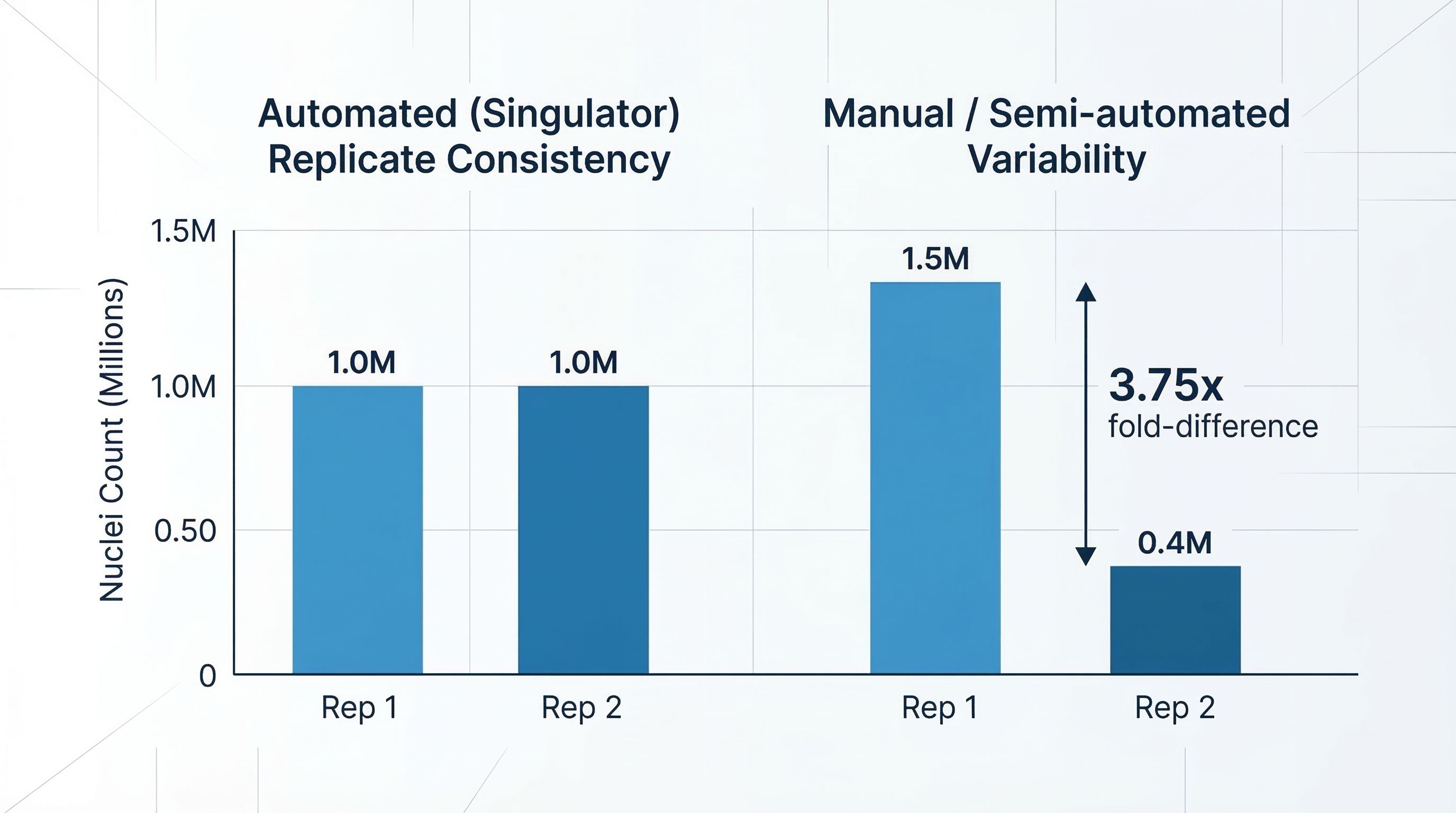
What consistency looks like in the data
In a study comparing the Singulator 200+ to semi-automated approaches for FFPE tissue processing, replicate samples told a clear story. The Singulator produced 1.0 million nuclei from the first replicate and 1.0 million from the second. The semi-automated method produced 1.5 million from one replicate and 0.4 million from the other, a 3.75-fold difference. Same tissue type. Same day. Different levels of control over the processing.
For a single Alzheimer's brain section, that inconsistency is academic trivia. For a longitudinal cohort processed over months by different technicians, or a multi-site consortium comparing results across institutions, it is the difference between publishable data and noise.
The contamination reduction
Erythrocyte contamination dropped from 5% with semi-automated methods to 1% with the Singulator 200+. Every contaminating red blood cell in a nuclei suspension wastes a sequencing barcode that could have captured a neuron, an astrocyte, or a microglial cell. Cleaner input means more of your sequencing budget goes to the biology you are studying.
Preserving the witnesses
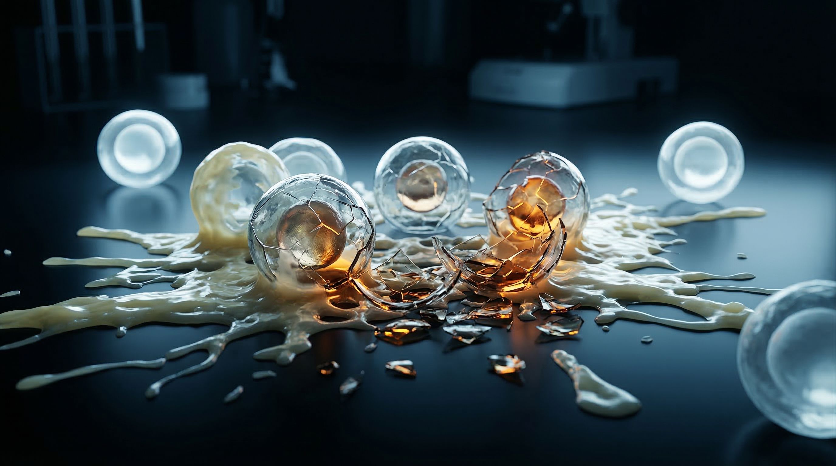
Recall from earlier in this series that the biggest problem with manual brain tissue processing is not speed. It is bias. Harsh trituration kills the fragile cells, the neurons and the delicate glial populations, while robust immune cells survive. The extracted population does not represent what was in the tissue. The witnesses you most need to hear from are the ones destroyed by the extraction.
The Singulator 200+ uses controlled mechanical and enzymatic processing within the sealed cartridge system. The forces are calibrated. The enzymatic conditions are standardized. The built-in filters handle the myelin debris that, in manual protocols, clogs equipment and contaminates samples. The result is a nuclei population that preserves cell-type diversity, keeping fragile populations intact rather than selecting for the toughest survivors.
This is what a forensic lab does. It preserves the evidence that fieldwork destroys.
From one section to data
The input requirements match the reality of biobank allocations. A single 35-micrometer FFPE section, or as little as 2 milligrams of tissue, is enough for the Singulator 200+ to produce over a million sequencing-ready nuclei. For researchers whose biobank allocation is measured in individual sections, this means the evidence from a single section can populate an entire single-nuclei sequencing experiment.
The nuclei are compatible with 10x Genomics Flex (probe-based chemistry designed for the fragmented RNA in FFPE tissue) and methods like PERFF-seq for rare cell capture. The snRNA-seq data from these nuclei also serves as companion data to inform Xenium spatial transcriptomics on adjacent tissue sections. A forensic lab does not dictate what analysis you run on the evidence. It gives you clean, well-preserved material and lets your science determine the next step.
The chain of custody, summarized
FFPE brain section loaded into the GREEN cartridge. Automated deparaffinization, no xylene, no fume hood. Transfer to YELLOW NIC+ cartridge. Automated nuclei isolation with debris filtration. Less than 5 minutes hands-on. Over 1 million nuclei. Ready for sequencing, spatial analysis, or both. The evidence has been processed. The chain of custody is intact.
In Part 5 of Cold Case, the cases are solved. Researchers pair spatial data with single-nuclei sequencing from adjacent sections, building brain cell atlases from tissue that sat in paraffin for decades. The evidence is finally testifying.