 BlogPart 5
Cold Case - Part 5: Case Closed
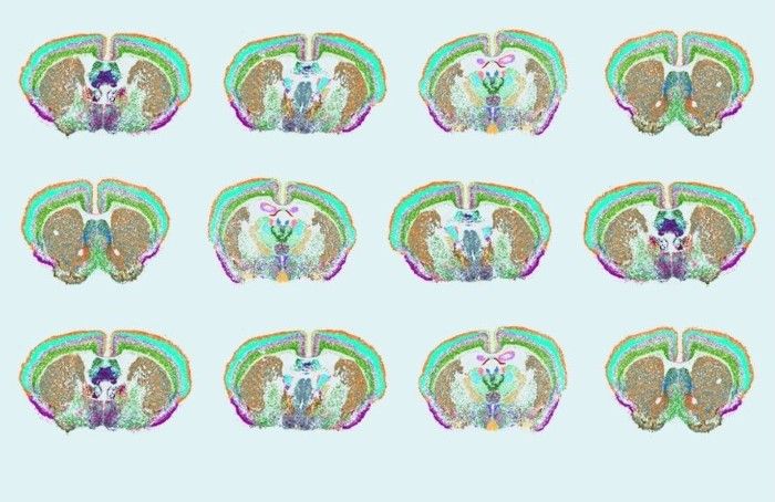
Brain atlases are being built. Archival tissue is finally talking. How standardized nuclei extraction and platform-agnostic analysis are solving neuroscience's oldest cold cases.
 BlogPart 5
Brain atlases are being built. Archival tissue is finally talking. How standardized nuclei extraction and platform-agnostic analysis are solving neuroscience's oldest cold cases.
 BlogPart 2
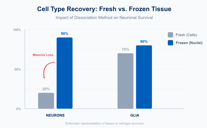
Manual extraction of nuclei from FFPE brain tissue destroys 50 to 60 percent of starting material. Fragile neurons die first, leaving biased results. Here is what goes wrong.
 BlogPart 1
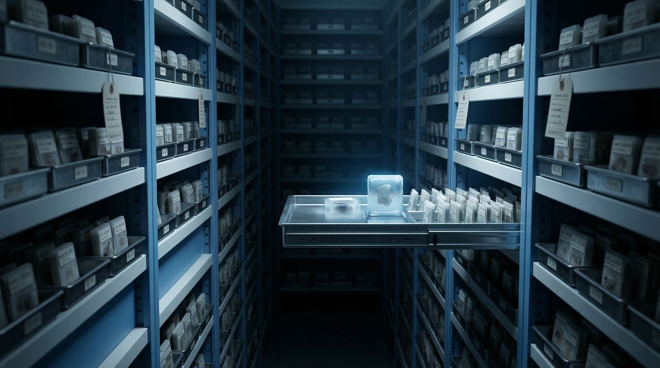
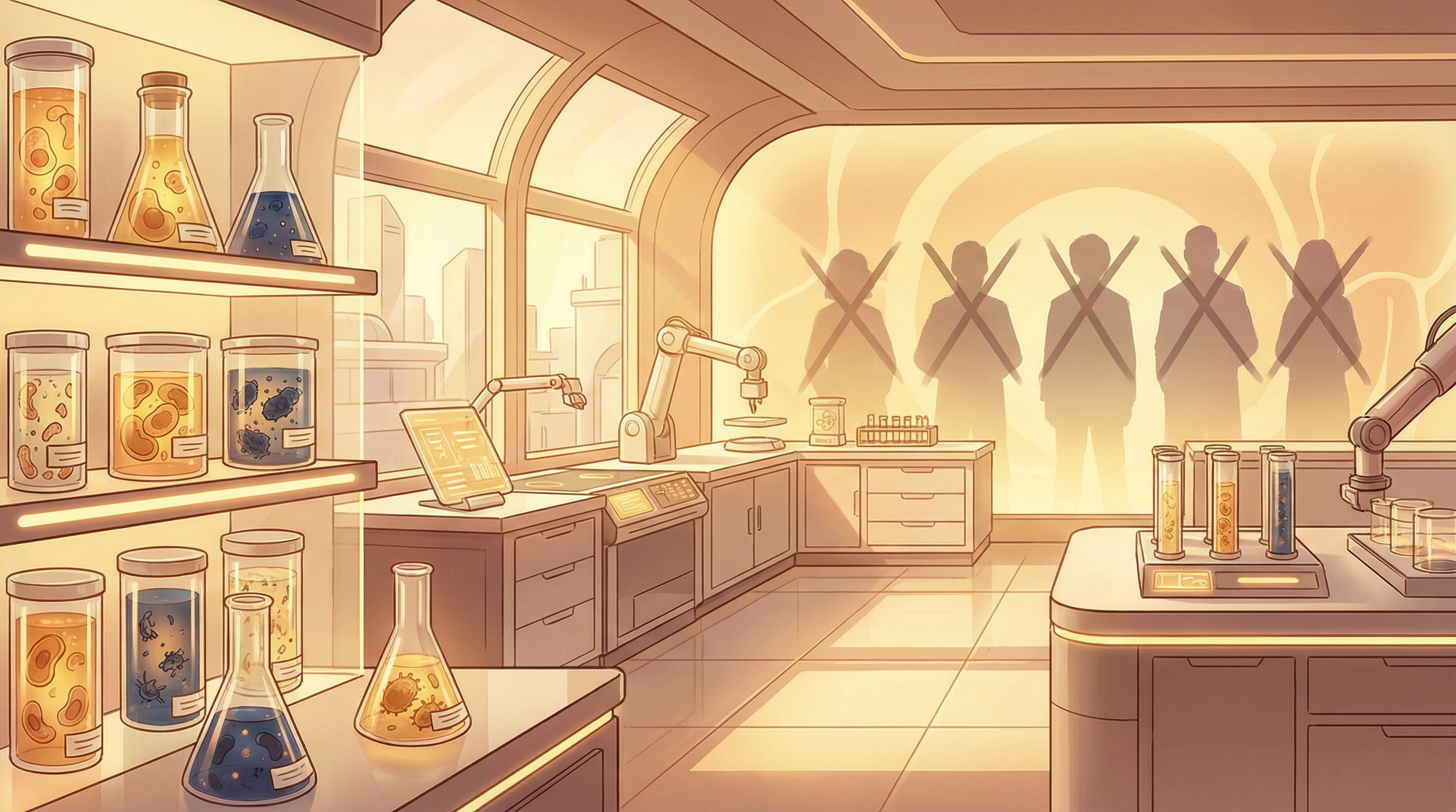
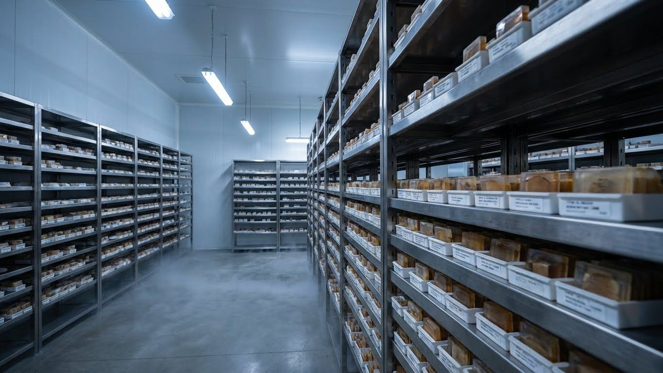
Millions of FFPE brain tissue blocks sit in biobanks worldwide, holding decades of evidence about Alzheimer's, Parkinson's, and neurodegenerative diseases. What if we could reopen these cases?
 BlogPart 4
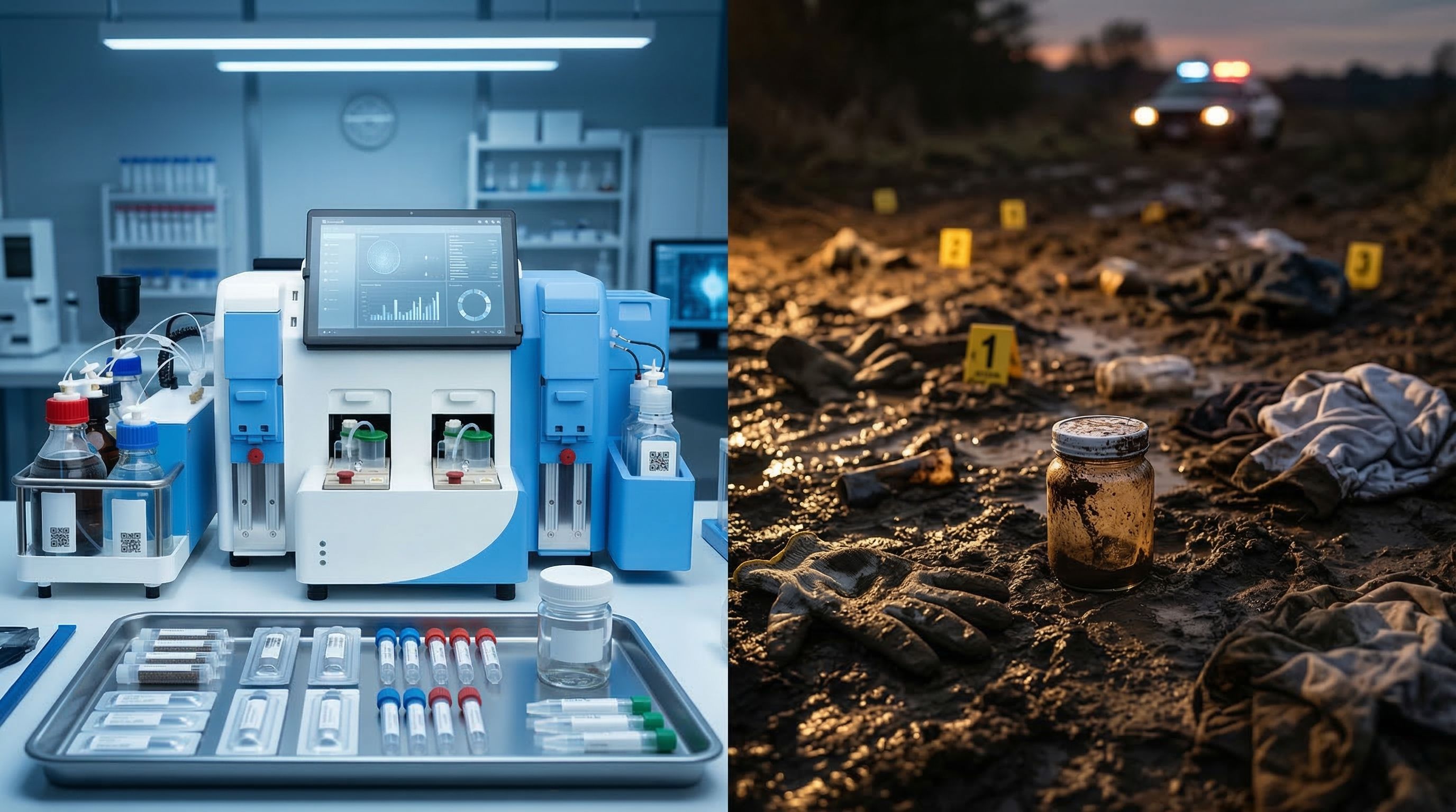
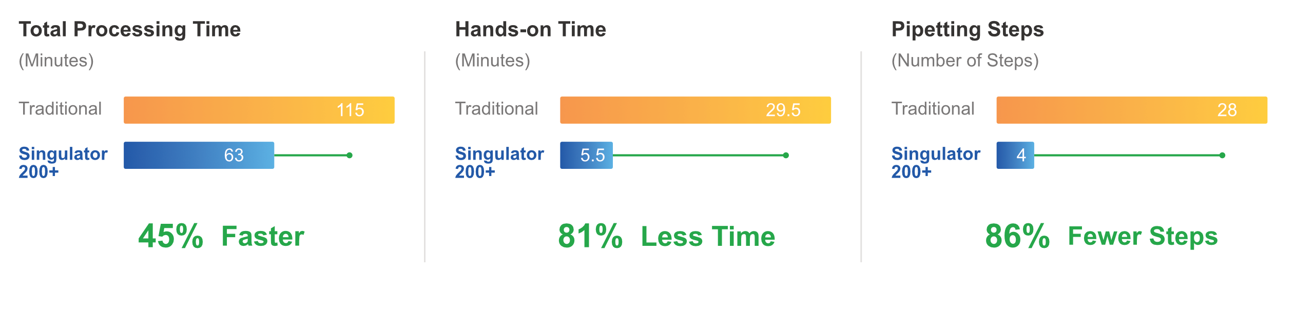
Manual FFPE processing destroys fragile neuronal nuclei and produces variable results. The Singulator 200+ automates the workflow with a two-cartridge system that delivers consistent, operator-independent results from irreplaceable brain tissue.
 BlogPart 3
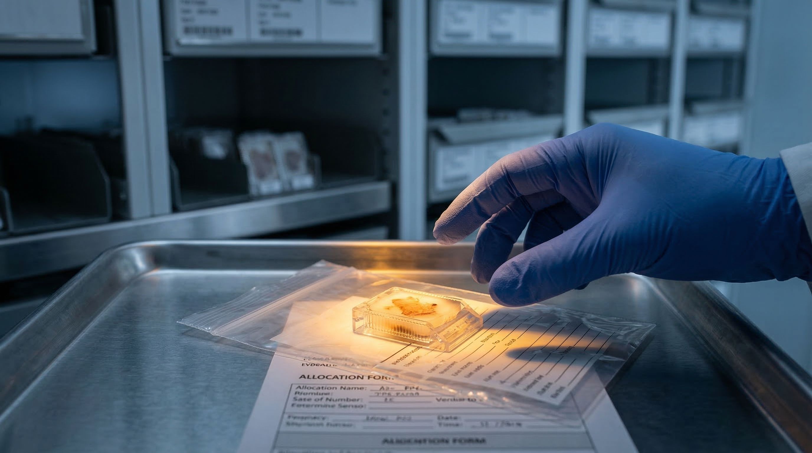
NIH biobanks give you one allocation of irreplaceable brain tissue. Manual processing destroys 50-60% before analysis begins. The extraction method is the real variable.
 BlogPart 1
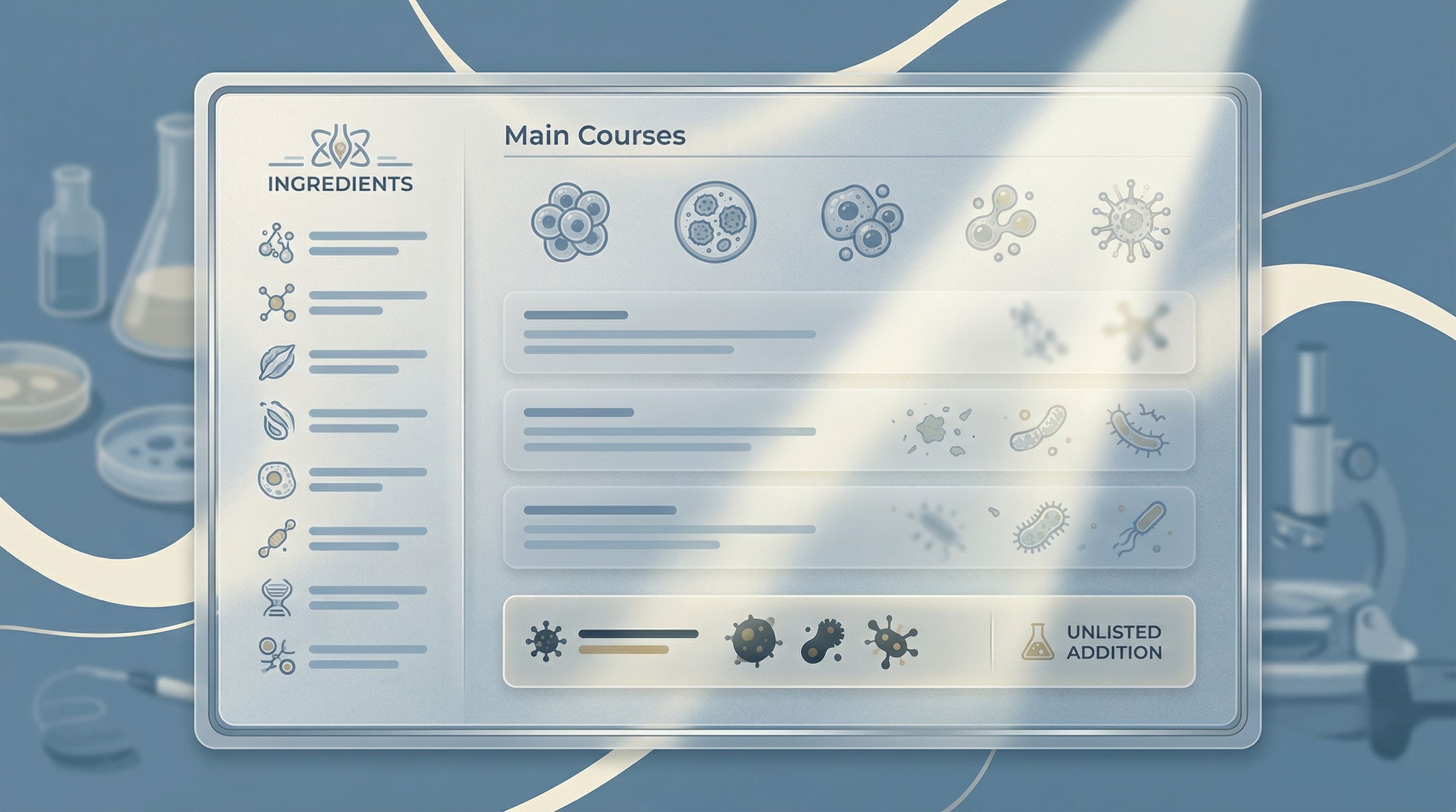
A cell count without debris quantification is like a menu without an ingredient list. Without knowing what invisible contaminants are present, every downstream decision becomes a gamble.
 BlogPart 2
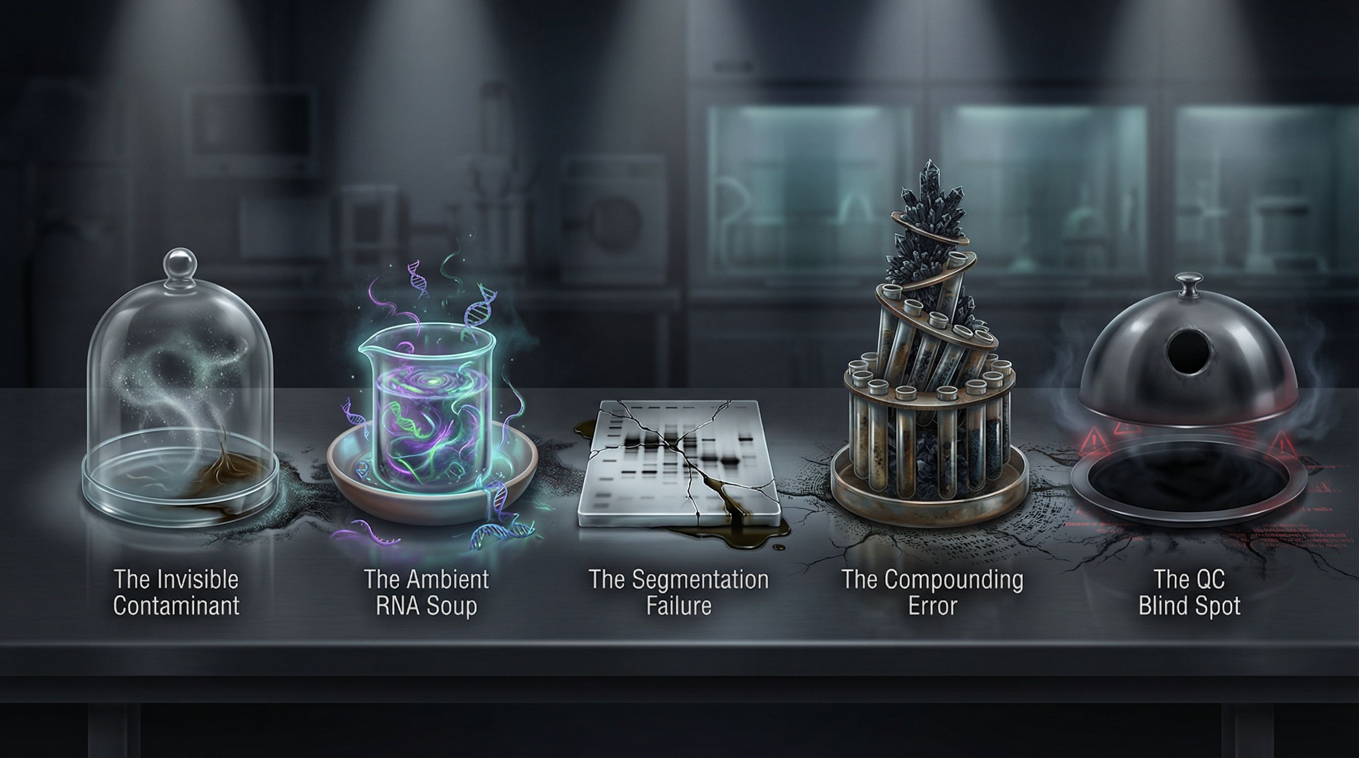
Five contaminants corrupt every sample. Image counters exclude them from counts but never reveal their presence. Until you can quantify what's actually in your sample—not just how many cells—these villains control the menu.
 BlogPart 3
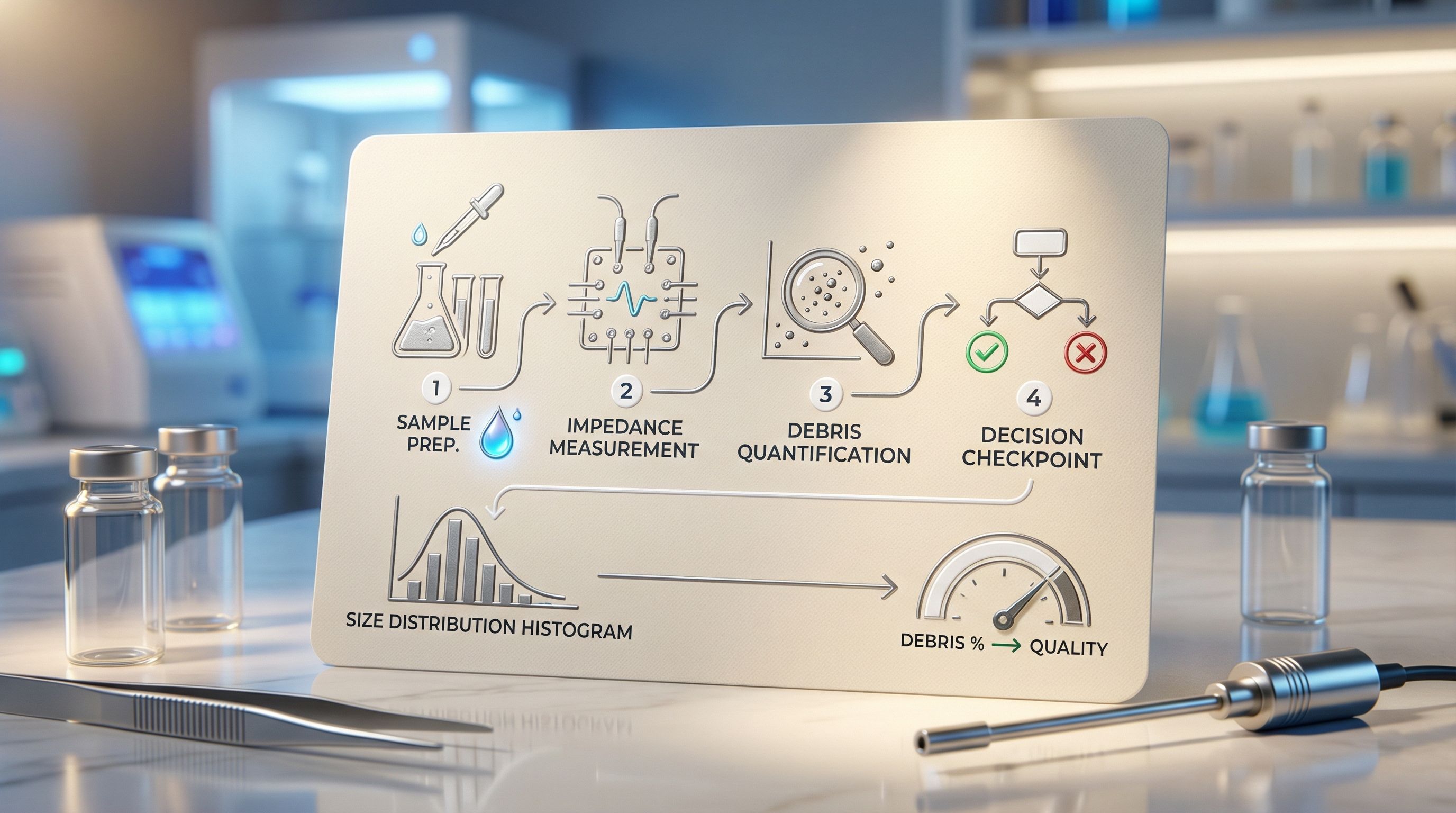
Better AI won't save your data. More training images won't expose the contaminants. Faster cameras won't quantify your debris. Physics will. The Coulter principle—the same physics that transformed clinical hematology—offers research laboratories what imaging never can: direct measurement of what's actually in your sample.
 BlogPart 4
The recipe for defeating the five villains isn't better algorithms or faster cameras. It's physics-based measurement that reveals what image counters hide: the complete composition of your sample. Direct size measurement. Complete population visualization. Standardized thresholds. Informed decisions. That's the recipe.
 BlogPart 5
Every laboratory has an invisible menu. Hidden ingredients contaminate samples. Villains corrupt data. Resources get wasted on samples that should have been cleaned up first. The question isn't whether these problems exist—they do, in every laboratory that relies on image-based counting alone. The question is whether you'll continue ordering blind, or finally demand to read the full ingredient list. Physics-based debris quantification isn't just an alternative to image counting. It's the missing QC checkpoint that transforms sample preparation from guesswork to measurement. From hope to confidence. From invisible menus to clean kitchens. The recipe is proven. The villains are defeated. The kitchen can be clean.
 BlogPart 1
Somewhere in your institution, there's a dusty shelf. Sitting on it: thousands of tissue blocks. Each one holds decades of clinical history. Patient outcomes. Treatment responses. Disease progression. Data that took years to collect. And can you blame researchers for walking past it?
 BlogPart 2
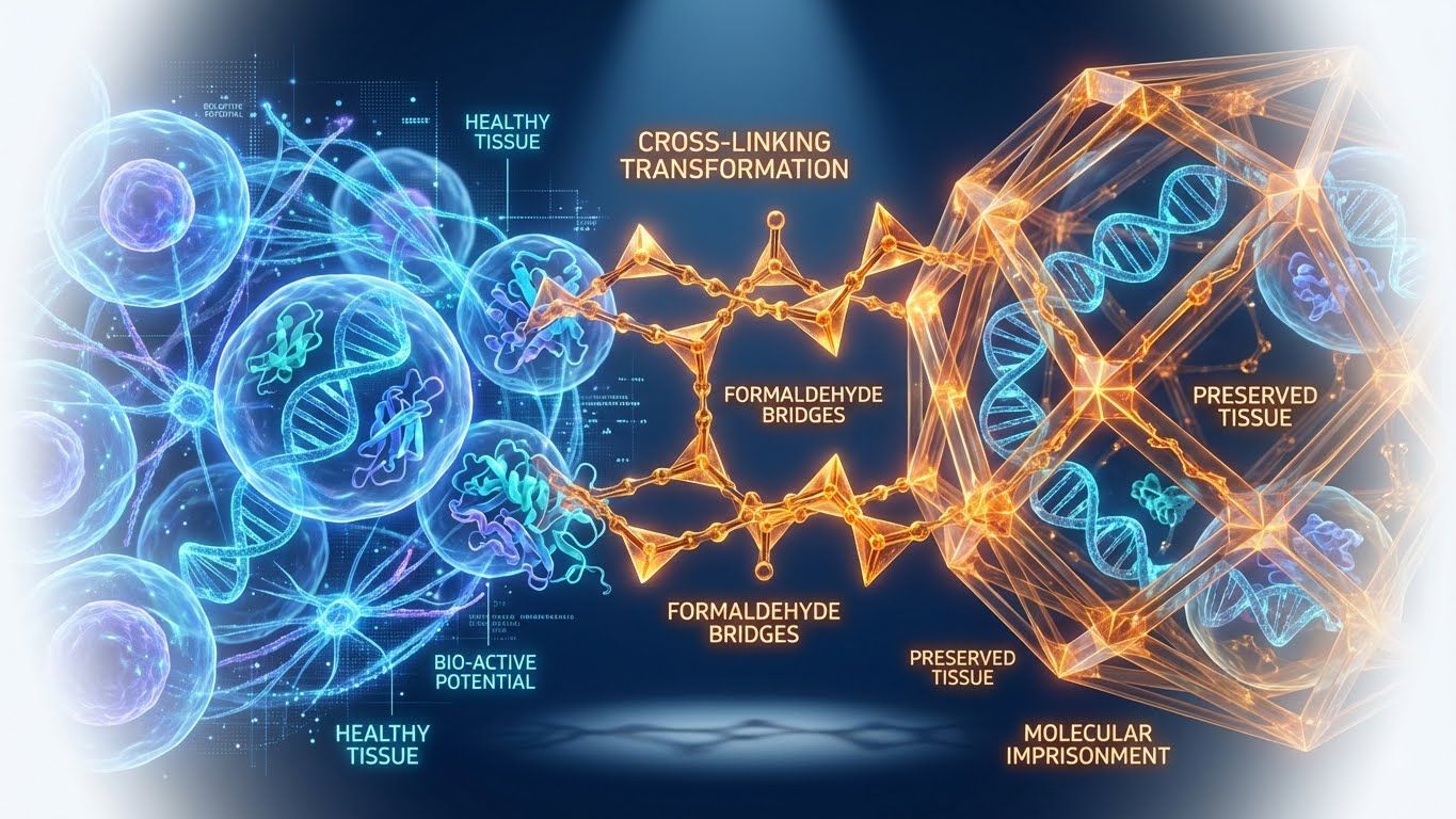
Here's a weird fact: the same chemical that saves your sample also traps everything useful inside it. Think about laminating a document. Great for protection. Terrible if you need to edit what's inside. That's formalin. And for decades, nobody could undo the damage.
 BlogPart 3
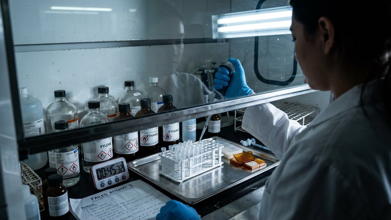
Third try. Third failure. The fume hood is running. The xylene smells terrible. And the protocol that worked last Tuesday is giving nothing but debris. Sound familiar? Here's the question nobody was asking: Is it your technique—or is it the physics?
 BlogPart 4
What if that two-hour protocol took sixty minutes? What if twelve pipetting steps dropped to four? What if the fume hood became optional? These aren't hypotheticals. This is what purpose-built automation looks like. And the difference isn't just time.
 BlogPart 5
Behind every tissue block is a patient who said yes. Yes to collection. Yes to research. Yes to the hope that their gift might help someone else. They trusted the science. The question now: are the protocols worthy of that trust?
 BlogPart 3
Fresh vs. Frozen: Which Side Are You On? Description: The debate between fresh tissue (whole cells) and frozen tissue (nuclei) divides labs. We explore why fresh dissociation often creates a "map of a disaster" through stress artifacts, and why frozen nuclei offer the unbiased, stable truth required for atlas-scale science.
 BlogPart 4
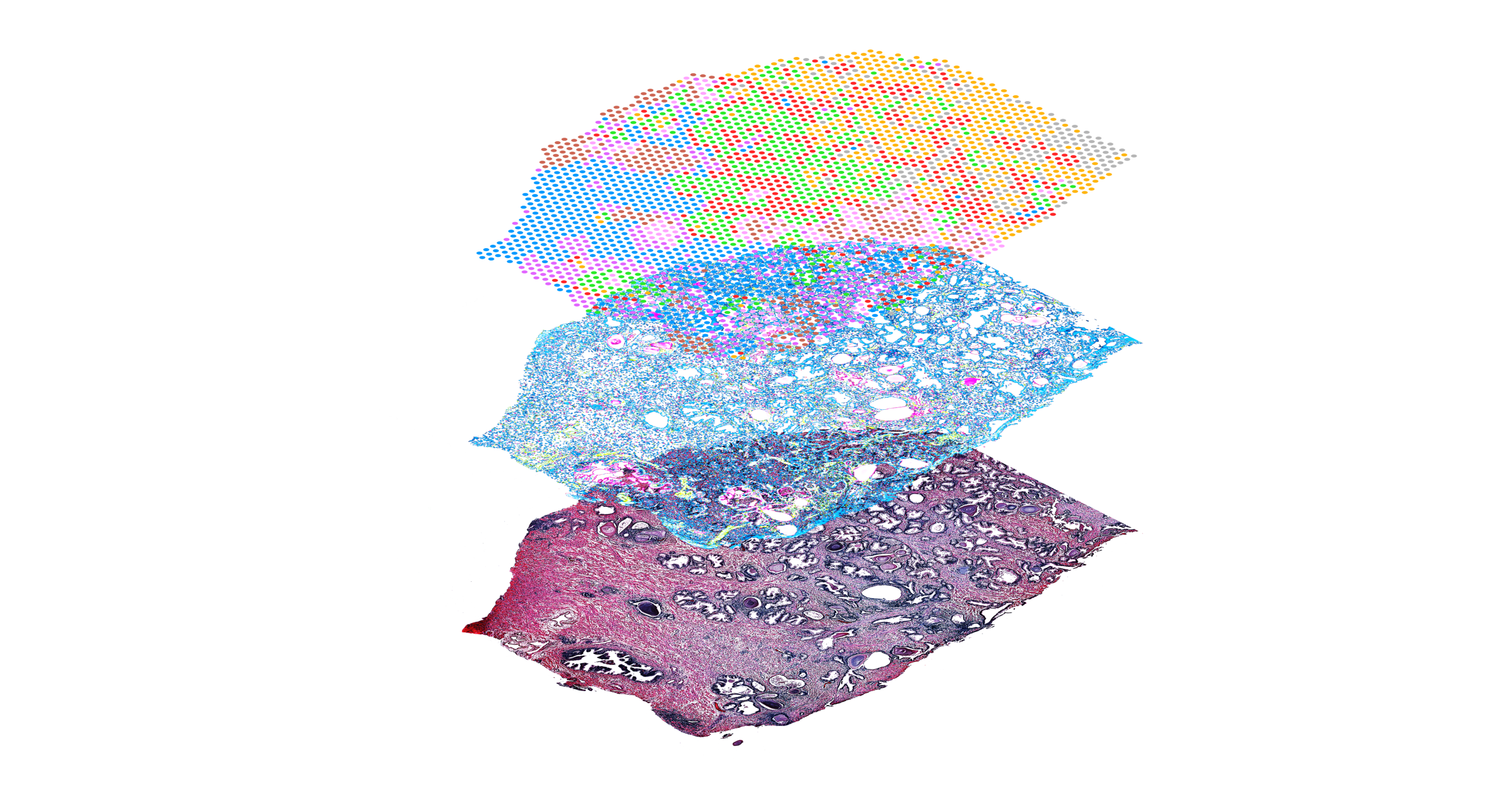
The List is Good, But Is the Map Better? Description: Single-nucleus sequencing gives you the "List" of cell types, but Spatial Transcriptomics gives you the "Map." Discover how combining these technologies creates a high-resolution "Precision Point," and how one automated platform can serve as the engine for both workflows.
 BlogPart 5
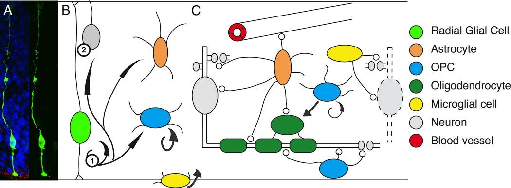
From the hidden world of glia to the final 3D atlas, the journey of modern neuroscience relies on one foundational step: sample preparation. We conclude our series by challenging researchers to prioritize clean, reproducible input to build the definitive map of the human brain.
 BlogPart 1
For a century, neuroscience focused exclusively on the neuron, dismissing glia as mere "packing peanuts." Discover how single-nucleus sequencing revealed the active, critical role of the brain's immune and support systems—and why this shift changes everything for Alzheimer's research.
 BlogPart 2
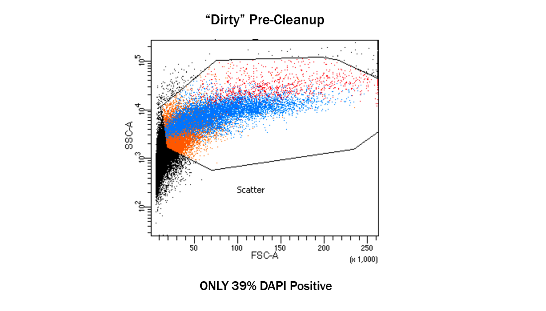
What is Ruining Your Frozen Experiments? Description: Every great story needs a villain. In frozen brain tissue processing, that villain is myelin debris. Learn how lipid contamination clogs microfluidics and ruins data, and see how the Singulator’s automated Protocol DP0006 neutralizes this threat to unlock biobank archives.
Our scientific team can help you match the right instrument and workflow to your research.
Contact Us